
The first cancer or tumor cell lines was HeLa that was derived from cervical cancer cells taken from Henrietta Lacks in 1951. In the early 1990s, a drug screening approach based on a “disease-oriented” program with a panel of 60 human cancer cell lines derived from nine different types of cancer (brain, colon, leukemia, lung, melanoma, ovarian, renal, breast, and prostate) was introduced by the National Cancer Institute (NCI; Bethesda, MD). This was aimed at facilitating high-throughput screening of a multitude of drug molecules for further studies using xenograft models. Research shows that 12 human cell lines as employed routine preliminary in vivo screens before using xenograft assays that are labor-intensive and more time-consuming (http://dtp.nci.nih.gov/branches/btb/hfa.html).
In another 2012-published report (Barretina et al, 2012), the Cancer Cell Line Encyclopedia showed that the copy number changes, mRNA expression and mutations were similar between the sequencing data and microarray expression profiles of 947 human cancer cell line with the data of primary tumors from publicly available databases. The conclusion of the study was that the cell lines “may provide representative genetic proxies for primary tumors in many cancer types.”
The testing of toxicity of molecules in animal models can be pursued only after the potential drugs show effects in appropriate in vitro cell line tests. This can support the notion of the 3 “R”s in the use of animal models:
- Replace the use of animals with alternative techniques.
- Reduce the number of animals used.
- Refining the approaches to allow minimum suffering to the animals.
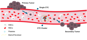
An important aspect of tumor formation is the microenvironment mainly composed of immune cells such as macrophages and fibroblasts that determines the formation and progress of tumor growth. The simulation of this environment by coculturing tumor-derived cell lines with fibroblasts or macrophages can allow the assessment of each cell and its effects along with the effects of effectors such as cytokines and lymphokines to understand carcinogenesis more in detail.
The use of 3D matrices such as Matrigel allows the development of models that closely resemble the tumor of their origin. This can allow the study of gene expression profiles, morphology, proliferation and drug resistance to hence serve as in vitro drug screening to predict human drug responses.
The knocking in and out of candidate genes in cell lines can allow studying the drug responses in detail. The use of genome-editing technologies such as CRISPR (clustered regularly interspaced short palindromic repeats) and TALENs (transcription activator-like effector nuclease) can easily allow the inactivation of genes or knocking out studies efficiently.
The application of “Omics” studies allow for the identification of molecular features that are common to various cancer types. Such common features account for the epithelial and mesenchymal origins for the tumors as demonstrated in breast and bladder cancers (Goodspeed et al, 2016).
Tumor cell lines shall remain a viable tool to continue to offer insights into one of the most dreaded conditions: cancer!
References:
Gillet, J. P., Varma, S., & Gottesman, M. M. (2013). The clinical relevance of cancer cell lines. Journal of the National Cancer Institute, 105(7), 452–458. https://doi.org/10.1093/jnci/djt007
Barretina J, Caponigro G, Stransky N, et al. The Cancer Cell Line Encyclopedia enables predictive modelling of anticancer drug sensitivity. Nature 2012;483:603–7.
Jennifer L. Wilding and Walter F. Bodmer. Cancer Cell Lines for Drug Discovery and Development.
DOI: 10.1158/0008-5472.CAN-13-2971 Published May 2014.
Andrew Goodspeed, Laura M. Heiser, Joe W. Gray and James C. Costello. Tumor-Derived Cell Lines as Molecular Models of Cancer Pharmacogenomics. DOI: 10.1158/1541-7786.MCR-15-0189 Published January 2016
