
Lungs are the vital part of the respiratory system for oxygenating blood and removing carbon dioxide. Their cellular composition includes immune system, epithelium and endothelium. The epithelium has direct contact with the external environment, thus driving the immune response. Its implications in respiratory diseases such as fibrosis, cancer, asthma, etc., have spurred substantial research. Mouse models have been suitable for in vivo studies to explore the mechanisms behind lung pathology. Therefore, research on mouse lung epithelial cells has surged in the last few years.
Mouse Lung Epithelial Cells
Appropriate research models for lung studies are available with mice. Their inbred strains offer genetic homogeneity. Their varied susceptibilities to lung diseases enable the investigation of underlying pathogenic mechanisms. They are also suitable platforms for translatable research since their pulmonary disorders are similar to those of people. The ability of genetic engineering in mice has also elevated its utilization as a replacement for humans in scientific studies. Therefore, mouse lung epithelial cells have gained preference for in vitro investigation.
Types of Epithelial Cells
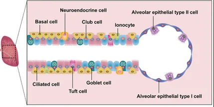
The epithelium lines the different regions of the lungs and vary substantially in its roles and structure. Almost 40 different cell types have been described in the lungs. Therefore, epithelium has been classified into three distinct regions- tracheobronchial or proximal airway, distal bronchioles, and alveoli. For instance, the epithelium of the tracheobronchial region comprises ciliated, club, basal, neuroendocrine, tuft, ionocytes, and goblet cell types. Bronchioles have a more ciliated and club cell population. Alveoli have flat alveolar type I and cuboidal alveolar type II pneumocytes.
Alveolar Epithelial Cells:
- Type I alveolar pneumocytes support the alveoli’s structure (Fig. 1). They contain xenobiotic detoxification enzymes, given their exposure to blood and gases. These enzymes can metabolize the toxins and prevent damage.
- Type II alveolar pneumocytes also contain Phase I and Phase II xenobiotic-metabolizing enzymes (Fig. 1). They also produce surfactant that prevents alveolar collapse. They show stemness by proliferating and replacing type I pneumocytes in cases of injury.
Tracheobronchial Epithelial Cells:
- Basal cells have cuboidal morphology (Fig. 1). They express markers- cytokeratins and Trp-63 and attach to a basement membrane. They can also differentiate into different epithelial cell populations, thus maintaining pulmonary homeostasis.
- Ciliated cells are columnar, possessing cilia on their luminal surface that beat in a wave motion to push the unwanted particles upwards and out of the system (Fig. 1).
- Clara or club cells differentiate and replace the ciliated cell type after their death (Fig. 1). Their level of xenobiotic-metabolizing is highest in the body, even more than in the liver.
- Goblet cells are mucus-producing cell types with a goblet appearance.
- Neuroendocrine cells are innervated cell populations present in low amounts (Fig. 1). They mediate communication between the nervous and immune systems by secreting neuropeptides. They are the first cell type to appear in murine pulmonary tissue and are crucial in fetal lung development.
Newly Discovered Epithelial Cells:
- Tuft cell, derived from basal cells, have microvilli and perform chemosensory, neuronal, and immunological functions (Fig. 1).
- Ionocytes are basal cell-derived cell types that express high amounts of CFTR protein, which is implicated in cystic fibrosis (Fig. 1).
Isolation of Mouse Lung Epithelial Cells
Enzymatic digestion is the ideal method to isolate the epithelial cells from the mouse lungs.
Extraction of Tissue Sample: Briefly, the protocol requires opening the thoracic cavity after sacrificing the mouse. The perfusion of the heart using a 25G needle flushes out erythrocytes. Afterwards, the lungs are removed and washed with a buffer. Mincing the lungs into small pieces with a sterile blade in an ice-cold buffer allows easy tissue digestion.
Enzymatic Digestion: The process entails the incubation of the tissue pieces in a collagenase enzyme solution for an hour. The larger tissue pieces settle down, whereas cells float in the upper supernatant. The removal of the supernatant every 15 minutes and replenishment with the enzyme solution provide a high cell quantity. After 4-5 repetitions, cell culture can proceed normally.
Cell Culture: The media for the cell culture is RPMI media, along with the use of fibronectin or collagen-coated plates. It yields two different adherent cell types- fibroblasts and epithelial cells. Fibroblasts are more susceptible to trypsin. Therefore, the cells that detach early from trypsin treatment can be removed to obtain only the epithelial cell population. Cell surface markers can characterize the purity of the population. Additionally, to isolate different cell types, options of magnetic-activated cell sorting (MACS) or flow cytometry-activated cell sorting (FACS) are available.
Lung Regeneration
The identification of diverse cell types in the lungs also led to the discovery of epithelial stem cells or progenitor populations and the pulmonary regeneration capacity. These progenitors maintain homeostasis in the lung by repopulating the dead cell population and playing a crucial role in injury repair. However, the notable part is that these progenitors vary according to their anatomical location. The tracheobronchial region contains a subpopulation of basal cells, the distal bronchioles have a subtype of club cells, and the alveoli contain Type II pneumocytes that show regeneration potential. The lineage tracing studies and in vitro experimentation have proven their stemness.
Additionally, scientists have recognized the markers of these subpopulations. Scientists have identified six different niches for these stem cells. This extraordinary capacity can prove advantageous in degenerative pulmonary diseases. Research has indicated that even the distinguishing stem cells and niches have the ability to cause different tumour types in the lungs.
Lung Regeneration in Disease and Therapy
Because of their role in acute lung injury, type II pneumocytes have been thoroughly investigated in epithelial regeneration. The repopulated cells have a different shape and produce scar tissue, which eventually impairs tissue function. Research has suggested that Notch and hypoxia signaling drive the impaired pulmonary regeneration, whereas Wnt signaling induces adequate functional regeneration. Sox protein seems vital to lung regeneration. A study demonstrated that Sox9 type II pneumocytes repaired lung injury and regulated immune responses, hinting at its use as a therapeutic target in the future. Early clinical trials have been employing bronchial basal cells in cell therapy for pulmonary disorders.
In a Nutshell
Pulmonary epithelium is essential for several tissue functions, especially regeneration. Lung damage after injury is attributed to the dysregulated regeneration process. Therefore, the pathways of regeneration are under exploration to develop suitable therapies. Although stem cell therapies have shown promise for pulmonary regeneration, many trials have included bronchial basal cells for treatment. Single-cell RNA sequencing studies have been trying to identify the markers of different epithelial cell populations with a focus on progenitors. Kosheeka provides the mouse lung epithelial cells with assured viability and purity so you can advance your research and pave the way for breakthroughs.
FAQs
Q: What are the functions of the lung epithelium?
It is involved in gaseous exchange, mucociliary clearance, xenobiotic detoxification, immune response against pathogens, regeneration, etc.
Q: Why use mouse pulmonary epithelial cell in research?
Mouse models offer the genetic homogeneity as well as ease of genetic editing, facilitating pulmonary research. Additionally, different models have different susceptibilities to lung disorders, aiding in decoding the pathological mechanisms. Therefore, their cells can provide more reliable insights.
Q: Which pulmonary epithelial cells exhibit stemness?
Basal cells, club cells, and Type II pneumocytes exhibit stemness by differentiating into various cell populations of epithelium.
Q: Which medium is used to culture mouse pulmonary epithelial cells?
RPMI is the preferred medium, along with collagen or fibronectin-coated plates, for in vitro culture.

