
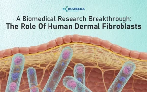
Human umbilical vein endothelial cells (HUVECs) or cord-derived endothelial cells that line the umbilical vein and serve as a suitable in vitro model for endothelial or vascular research. Endothelial cells constitute blood vessels, lining them on the luminal side and creating a semi-permeable barrier. Initially, their barrier properties, developmental process, and permeability have been under investigation. However, research on HUVECs has revealed more functions of the endothelium than previously thought. These studies have also changed the ideology that the endothelium is merely a static layer into accepting the dynamic nature of the tissue. The research on the role of endothelial cells in vascular disorders, physiological processes, and their upcoming advances in cell biology has been growing.
Characterization
The endothelial cell research began with a research paper. In 1970, Jaffe et al. achieved a milestone by isolating and culturing Human Umbilical Vein Endothelial Cells in the in vitro environment. HUVECs became the first in vitro models for vascular research, contributing substantially to endothelial studies. Their ease of harvesting promoted their extraction and in vitro applications. Currently, most of the knowledge in vascular biology- signaling pathways, cellular response, genetic profile, etc. has been derived from studies on HUVECs.
Such studies have also evolved the culture and characterization of HUVECs. Initially, they were recognized as cobblestone-shaped cells with the presence of von Willebrand factor (vWF) and Weibel Palade bodies. However, with time, their marker panel expanded to include CD31, CD144, CD34, CD62E, CD62P, CD106, CD54, etc. Assays such as dil-Ac-LDl to evaluate uptake of lipoproteins and the Matrigel assay have also emerged for analyzing the functional potential of these cells.
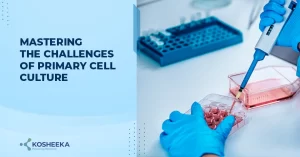
Tips for Culture
Culturing HUVECs has not been exactly a walk in the park. Two factors that have always confounded the researchers- an appropriate medium for cells and cell adhesion to the surface of a flask or dish.
Medium: Nowadays, commercially available HUVECs are supplied with an option for a suitable medium. It has somewhat lessened the requirement for media optimization and formulation from the end of researchers. However, some might choose to formulate media based on different applications. M199 is considered to be an ideal medium for HUVECs and endothelial cells in general. MCDB131 with high magnesium also stimulates endothelial cell proliferation. Some labs also employ DMEM, but it is not suitable for HUVEC culture. Concentration of fetal bovine serum (FBS) might need to be high in the initial isolation and culture period. However, 10% FBS generally suffices the optimal cell growth. Medium is supplemented with growth factors like vascular endothelial growth factor (VEGF) and heparin. Both promote cell proliferation. Heparin facilitates cell adhesion, intercellular communications, and enhances the effects of endothelial cell growth factor (ECGF).
Coating: Experimentation with HUVEC culture has shown that cell adhesion is vital for cell survival and proliferation. Although many times, growth of HUVECs has been noticed on a cell culture dish, coating is still recommended for cells. Coating the plates or flask with gelatin or fibronectin has demonstrated prolong cell survival.
Research on Human Umbilical Vein Endothelial Cells
Drug testing:
The rapid advancements in nanotechnology and bioinformatics have accelerated the drug discovery and development process. The evaluation of their toxic effects has expanded from screening only on hepatocytes to the inclusion of other tissue-specific cells. Drugs can alter the endothelium and result in disorders such as thrombosis and atherosclerosis. It can have severe long-term effects with subsequent drug withdrawal from the market. Therefore, drug effects are assessed on HUVECs to account for any vessel-associated complications.
Angiogenesis:
Angiogenesis, or the process of blood vessel formation, is a key event in the morphogenesis of vessels and injury repair. Cells use either sprouting or splitting angiogenesis. Sprouting angiogenesis entails the proliferation and migration of endothelial cells to create a sprout. The tip cells guide the other cells under stimulation by VEGF. In splitting or intussusceptive angiogenesis, the vessel wall extends towards the lumen and fuses with the opposite site to split the vessel into two. Knowledge of the mechanisms behind these processes is insufficient thereby promoting deep exploration into the impact of factors such as hypoxia, VEGF concentrations, etc., with a focus on its applications in vessel development and pathogenesis.
Physiological Processes:
The Recent few decades have divulged that the endothelium can modulate its phenotype in accordance with the environmental stimuli. It maintains vascular homeostasis by retaining an anti-coagulative and anti-thrombotic surface. However, vessel injury can trigger its transformation into a coagulative surface. Several molecules, such as PAI, t-PA, u-PA, thromboxane, NO, etc., maintain the delicate balance between the two phenotypes. Nitric oxide (NO) is a key signaling mediator for controlling vessel diameter. With implications of these processes in diseases, research on HUVECs have considerably increased.
Barrier Property:
The primary function of the endothelium is to govern the traffic across tissue. Its permeability varies in different tissues. A key example of its barrier function is the blood-brain barrier. Endothelial cells are held together with tight junctions, rendering the barrier impermeable. Scientists have therefore been studying the details of the endothelial permeability, migration, and junctions to increase the delivery of drugs across the barrier.
Oncology:
Blood vessels supply nutrients to the tumor, promoting its growth. It has urged the development of anti-angiogenic therapies for cancer treatment. These therapies has also paved the way for nanomedicine in cancer. Nanomedicine exploits the disrupted nature of tumor vasculature and small size of drugs to increase the distribution of drugs into the tumor tissue while minimizing off-target distribution and adverse effects. Therefore, tumor treatment plan have integrated anti-VEGF antibodies such as bevacizumab to inhibit vessel growth in tumor tissue.
Inflammation:
Chronic inflammation has been a significant process behind several disorders or their worsening. It involves the migration of immune cells to the site of injury or infection. Migration and transport across the vessels require endothelial signaling. Endothelial cells release chemokines that aid in immune cell recruitment. They also upregulate adhesion molecules to support cell attachment to the endothelial surface and their migration into the tissue. Migration process employs another set of molecules and signaling molecules to retract their junctions and allow cells to pass across the layer.
Product-Related Queries, Or Partnership Inquiries
Future Perspectives
HUVEC have significantly propelled the endothelial research and the drug discovery process for vascular disorders. Since the beginning of in vitro study in endothelium, HUVECs have been the only in vitro tools for exploration. The recent studies have been demonstrating the variation among different types of endothelial cells, prompting their isolation and utilization in cell culture studies. However, many of the processes delineated on HUVECs remain the same across the different cell types. Furthermore, Human Umbilical Vein Endothelial Cells are more suitable due to their higher proliferation and ease of extraction. Their use in 3D tissue engineering has also increased. Vascularization of organoids with endothelial cells recapitulates the in vivo environment more accurately while prolonging the survival of organoids. To empower your research, Kosheeka avails ethically sourced and well-characterized HUVECs. Our team of experts is available to guide you with cell culture and troubleshoot any problem you may encounter.
FAQs:
Q – Can HUVECs grow on uncoated tissue culture dishes?
HUVECs have demonstrated adherence to uncoated culture dishes and showed optimal growth. However, coating of gelatin or fibronectin is recommended if cells do not attach to the culture dish.
Q – Is DMEM a suitable medium for HUVECs?
The ideal medium for HUVECs is M199 supplemented with FBS. However, many research studies have used low-glucose DMEM with 10% FBS and demonstrated proper cell growth.
Q – Should I add growth factors to the culture medium for HUVECs?
Serum is generally considered the source of growth factors for HUVECs. In case of serum-free medium addition of growth factors such as VEGF, bFGF, and heparin is recommended.
Q – What is the dil-Ac-LDL assay?
Dil-acetylated low-density lipoproteins assay incubates HUVECs with acetylated low-density lipoproteins. The lipoproteins are conjugated to a fluorescent probe containing 1,1’-dioctadecyl-3,3,3’,3’-tetradecylindocarbocyanine perchlorate (Dil). The cellular uptake of lipoproteins is measured by cell fluorescence. The assay distinguishes HUVECs from other cells by the endothelial cell capacity to internalize lipoproteins.
