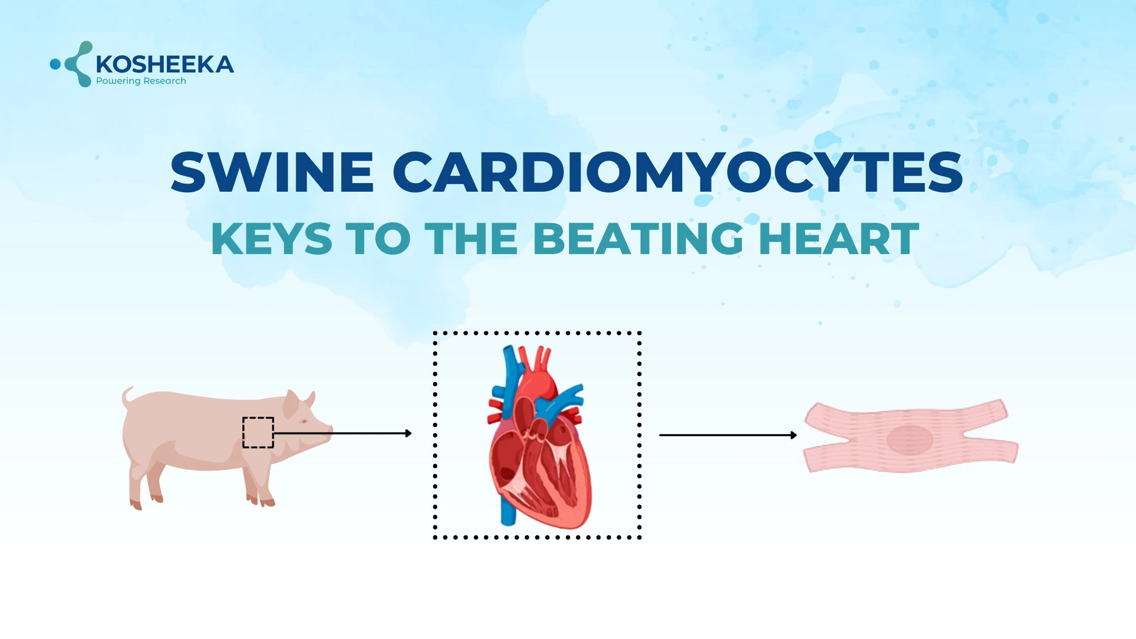
During development, the heart is the very first organ to take shape. Cardiomyocytes, or cardiac muscle cells, coordinate together for rhythmic beating that pumps blood to different tissues. Any damage to these cells or their dysfunction can cause severe disorders. Therefore, scientists have been working on these cells to gain knowledge about their underlying pathways and their involvement in disease to design effective therapies. The swine animal model has become the center for cardiovascular research due to sharing similarities with the human heart and physiology. Thus, research has accelerated on swine cardiomyocytes to recapitulate the in vitro findings with in vivo models.
Cardiomyocyte Structure
Cardiac muscle cells have a single nucleus in their center. The arrangement of their myofibrils and sarcomeres is visible as striations. Their plasma membrane or sarcolemma comprises voltage-gated ion channels for contractile activity. These cells contain transverse tubules (t-tubules) in the cytoplasm. They are invaginations of the membrane that position calcium ion channels in the vicinity of calcium-sensing and releasing channels. Cardiac muscle cells join via gap junctions and desmosomes, forming a branched structure with intercalated discs. Desmosomes anchor cells together while gap junctions permit the propagation of an action potential from one cell to another.
Cardiomyocytes differ from other muscle cells, like skeletal and smooth muscle cells, in various aspects. Cardiac muscle cells have shorter t-tubules that do not link to the endoplasmic reticulum and the presence of ion channels in the membrane. They are primarily aerobic and fulfill their requirement via large numbers of mitochondria and myoglobin reserves.
Types of Cardiomyocytes
Initially, cardiomyocytes were categorized into contractile and conductive components. Conductive or pacemaker cells present in Purkinje fibers have lower myofibrils and are specialized in conducting action potential more rapidly in comparison to the contractile phenotype. With the advent of technology, other distinguishing features surfaced apart from electrophysiological processes.
According to transcriptome data, two different populations of cardiomyocytes reside in the heart- ventricular and atrial. Both types of cardiomyocytes have distinct developmental processes and electrophysiological properties. Ventricular cardiomyocytes contain genes for sarcomere formation and stabilization, whereas atrial subtypes have genes for cell differentiation and mechanosensing.
Function of Cardiomyocytes
Cardiomyocytes are responsible for orchestrating the coordinated contraction of cells across the tissue. Sarcomere, consisting of myosin-actin filaments, is the functional unit of myocytes. T-tubules participate in signaling, action potential initiation, signal propagation, and excitation-contraction coupling (ECC). Several ion channels, such as voltage-gated L-type (long-lasting) Ca2+ channels, Na+/K+ ATPase, Na+/K+ exchanger, etc. The complete action potential can be divided into five different phases:
- Phase 1: The resting phase maintains a -90mV resting membrane potential via Na+/K+ channels, extruding three sodium ions for influx of two potassium ions.
- Phase 2: When the action potential reaches cells, fast Na+ channels open rapidly, causing an influx of sodium ions, leading to an increase in membrane potential to +52mV.
- Phase 3: Fast Na+ channels close instantly while Cl- and K+ channels open resulting in entry of chloride ions and potassium ion efflux initiating the repolarization process.
- Phase 4: Voltage-gated L-type (long-lasting) Ca2+ channels open leading to small increase in cytoplasmic Ca2+. It leads to activation of Ca2+ channels in sarcoplasmic reticulum further elevating cytoplasmic Ca2+ levels. High Ca2+ binds to troponin, dissociating tropomyosin from actin, which enables actin-myosin interaction. Myosin slides across the filament by ATP hydrolysis causing contraction. Meanwhile, potassium efflux balances calcium influx.
- Phase 5: High Ca2+ is sequestered in various manners- binding to calmodulin, influx of Ca2+ into sarcoplasmic reticulum by ATP-dependent SERCA-2 Ca2+ pump, and internalization by calsequestrin. Delayed K+ channels that activate at a later stage restore the membrane potential, transitioning to Phase 1.
Why Swine Cardiomyocytes?
Cardiomyocytes are attractive candidates to study cardiovascular functions. However, isolating them from humans is a challenging task, limiting the availability of human cardiomyocytes. Even cardiomyocytes derived from induced pluripotent stem cells were employed for studies, but they also lacked the maturation and structural features of human cells.
Swine animal models have been frequently used due to their compatibility with surgical interventions and gene editing. Swine heart has a comparable size to that of the human heart and also shows identical structural and electrophysiological characteristics to human cardiac tissue. Even their hemodynamic properties bear a close resemblance. Therefore, swine heart and its cardiomyocytes are more suited for in vitro research. Research has also revealed similarities in the genetic profiles, contractility, and heart rate between human and swine muscles. The parallels between both species have increased the use of swine cardiac muscle cells in research.
Product-Related Queries, Or Partnership Inquiries
Applications in Spheroids
Three-dimensional (3D) constructs of the heart represent the fundamental concept of tissue functioning. Cells require structural and environmental cues to sustain the genetic profile and replicate the functions similar to in vivo tissue. Cardiac spheroids and organoids have therefore gained popularity. These structures can aid in the development of more appropriate medications by offering precise information about the physiology and pathophysiology.
In myocardial infarct models, the implantation of cardiac spheroids made of cardiomyocytes has shown benefits. Different strategies are under investigation to improve the generation of spheroids, such as variation in biomaterials and integration of other cardiac cells. Based on this approach, scientists have developed a heart patch comprising myocytes and biomaterial to repair cardiac damage. The ongoing clinical trial of the product is determining the therapeutic potential of the product in humans with cardiac disorders.
Applications in Drug Development
The prevalence of death by cardiovascular disorders has been on the rise. Formulating better therapeutic alternatives is crucial. Therefore, many researchers have been working on swine cardiomyocytes to find appropriate targets for drug development. Furthermore, the new drugs are assessed on these cells to estimate their efficacy in the disease.
Advances such as in vitro culture, computational biology, and high-throughput screening have significantly progressed drug development. A vital part of the process is screening drugs for toxicity. Early studies focused on hepatocytes for the screening process. However, drugs that qualified the screening process have shown substantial cardio toxicity, eventually leading to their withdrawal from the market. Therefore, cardiomyocytes have found applications for evaluating drugs for adverse effects on cardiac tissue.
Conclusion
Cardiomyocytes are vital for cardiac contraction and function. These cells have been a constant subject of study for years. Researchers have obtained valuable insights from these cells. The low availability of human cardiac muscle cells has urged the search for a suitable alternate. Many studies have been conducted on mouse and rat models. However, the swine model surpasses them by comparable features of human and swine cardiomyocytes. They can unlock the mechanisms behind the disorder and repair them to produce a suitable medication. Scientists have been focus on cardiac reprogramming and generation of 3D constructs that have therapeutic applications. Kosheeka adds value to such scientific studies by delivering a pure population of cardiomyocytes. Its team of PhD experts ensures the viability of these cells and runs stringent tests for a contamination-free product.
FAQs:
Q – What is the function of cardiomyocytes?
Cardiomyocytes are cardiac muscle cells responsible for the contraction of the heart and for pumping blood throughout the body.
Q – What are the types of cardiomyocytes?
Cardiomyocytes have been broadly classified as contractile and conducting types based on their cell structure and function. However, transcriptome studies have found more subsets of these cells depending on their different location.
Q – Why are swine cardiomyocytes used in research?
Swine cardiomyocytes share similarities with human cardiomyocytes. Additionally, swine hearts show identical electrophysiological and hemodynamic properties. Thus, swine cardiomyocytes offer a better in vitro model for research.
Q – What are the applications of cardiomyocytes?
They are presently used for drug development and testing. Moreover, scientists are driving cardiac regeneration by creating 3D constructs from these cells.

