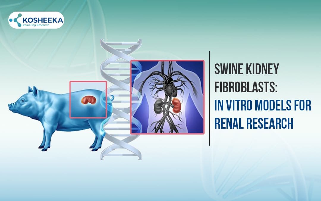
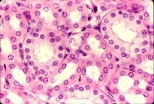
Swine kidney fibroblasts are reliable in vitro tools for renal research owing to the similarities between pigs and humans. Fibroblasts are essential for maintaining the renal structural framework by producing the extracellular matrix. Due to their role in repairing tissue injury and fibrosis, they have become a focal point of research. Therefore, research on swine kidney fibroblasts has focused on studying kidney functions, understanding renal disease mechanisms, and formulating innovative treatments.
Why Swine Kidney Fibroblasts?
Pigs share a long history of applications as animal models in biomedical research. The species have been especially investigated for xenotransplantation studies. They have genetic, anatomical, and physiological similarities with humans. Due to their large size, they are compatible for the development and testing of surgical and endovascular techniques.
The resemblance between pigs and humans has fueled the research on metabolic renal disorders in the porcine cellular models. Comparative genome analysis has also established the genotypic similarity between the two species. Furthermore, the swine genome sequence is available. It enables gene editing of swine models to develop human disease-specific models for in vitro and in vivo studies. These factors have contributed significantly to the rise of cultivating Swine Kidney Fibroblast Cells.
Isolation of Porcine Renal Fibroblasts
Utilization of either the explant technique or enzymatic digestion extracts pig kidney cells. Usually, needle core biopsy for kidney tissue slices or whole tissue extraction is employed for obtaining tissue samples. The buffer for sample storage is ice-cold phosphate buffer saline (PBS) containing antibiotics. At the time of isolation, tissue is washed and minced in a buffer, followed by washing in DMEM until the supernatant is clear.
Enzymatic Digestion
This method entails incubating tissue pieces in a collagenase II solution. It is essential to remember that a fresh enzyme solution in a buffer, pre-warmed at room temperature, offers better results. After incubation, filtration of cells through a strainer and washing will yield a population of swine kidney fibroblast cells.
Explant Method
Instead of enzymes, the explant method exploits cell migration from tissue to the culture plate. The approach spreads the sample on a gelatin-coated petri dish. Making crosswise scratches can aid in tissue attachment. Gentle addition of medium prevents the tissue from detaching. Within 10-14 days, cells from the tissue migrate to the petri dish. It is possible that cells show epithelial cell-like cobblestone morphology. However, later, they will assume the fingerprint appearance of fibroblasts. At this point, the cells can be subcultured.
Pig Renal Fibroblast Characterization
The cell population in renal tissue is diverse and complex. Therefore, characterization of fibroblasts after isolation is the final step to ensure purity of the population. Immunocytochemistry and immunofluorescent techniques typically measure the cell-specific markers, such as vimentin, α-smooth muscle actin (α-SMA), and collagen I. Additionally, analysis of the negative markers, such as vWF, VE-cadherin, CD34, etc., and pancytokeratin markers indicates the absence of other types of renal cells, such as epithelial and endothelial cells.
Porcine Kidney Fibroblast Culture
Dulbecco’s Modified Eagle Medium (DMEM) with 10% fetal bovine serum is ideal for Swine Kidney fibroblast proliferation. Changing the culture medium twice weekly helps sustain cell viability and growth. Exceeding 80–90% confluency increases the risk of cellular senescence or changes in cellular morphology, thus emphasizing subculture at the right confluency. Typically, the upper limit of passage number for swine renal fibroblasts ranges between 5 and 15 before an apparent decline in cell behavior and survival.
Swine Kidney Fibroblast Cell Lines
The induction of the human telomerase reverse transcriptase (hTERT) gene confers immortality to swine kidney fibroblast cells, making continuous or immortal cell lines. Without compromising the essential characteristics of fibroblasts, this method prolongs swine kidney fibroblast proliferation. For genetic and immunological research on the kidney biology of pigs, such immortalized cell lines provide a reliable and reproducible platform. This is especially relevant given the growing interest in pig-to-human xenotransplantation, where understanding swine immunogenicity is critical. Additionally, induction of pig TERT is also attainable through a transposon, a non-viral gene delivery method. Notable examples of swine kidney fibroblast cell lines include PK15, LLC-PK1, PFC, etc.
Swine Kidney Fibroblast in India
India is expanding its research on kidney diseases and transplant biology with increasing adoption of swine models in institutes. The ability to model renal diseases using swine cells are beneficial preclinical studies and clinical applications within India’s healthcare ecosystem. Given India’s increasing focus on advanced biotechnology and translational research, pig renal fibroblasts offer several advantages that are aligned with this progress.
Kosheeka is a vital part of this progress. It is a cell culture company that offers a wide inventory of primary cells and cell lines for scientific research. It saves researchers’ time in isolating and characterizing cells. The company’s standardized protocols confer uniformity in each batch of cells, reducing experimental variation with the use of different batches. It also offers customization services to extract cells from specific donor profiles, such as specific disease models, age, gender, etc.
Applications
Kidney Disease Modeling
Fibroblasts are a central player in fibrosis, which involves excessive deposition of an extracellular matrix that disrupts normal kidney architecture and function. By studying these cells in vitro or within swine models, scientists gain insight into how kidney injury progresses, the signaling pathways involved, and cellular responses driving fibrosis and inflammation.
Drug Testing
Porcine renal fibroblasts provide an in vitro system that enables researchers to assess drug toxicity and efficacy before moving to clinical trials. Toxicity screening on these cells identifies potential nephrotoxic effects earlier in drug development, reducing the risk of adverse effects in patients. It streamlines the drug development process and improves safety profiles for new treatments targeting renal diseases.
Xenotransplantation Research
Pig organs, including kidneys, are viewed as strong candidates for addressing the shortage of human donor organs in transplantation. Pig kidney fibroblasts are studied to understand immune responses to pig tissues, which is a critical barrier in cross-species transplantation, known as xenotransplantation. These fibroblasts express specific swine leukocyte antigens that can trigger immune rejection in humans. Investigation into these immune interactions promote the development of strategies to reduce immune rejection such as genetic modifications or immunosuppressive therapies.
Regenerative Medicine
Porcine kidney fibroblasts have demonstrated remarkable plasticity. Studies show that by introducing specific transcription factors, these fibroblasts can be directly reprogrammed into hepatocyte-like cells, mimicking liver cells with functional metabolism. This capacity for cellular conversion opens exciting possibilities for regenerative therapies not only confined to renal applications but also extending to other organ systems.
Conclusion
swine kidney fibroblasts enable the study of basic kidney biology and disease. Their applications have also extended to translational regenerative medicine. The immortal pig kidney fibroblast cell lines ease the culture process and facilitate long-term studies. Research on these cells will advance kidney disease treatment. India has considerably invested in renal research employing these cells. Kosheeka is supporting this endeavor with its high-quality swine kidney fibroblasts in India.
What makes swine kidney fibroblasts ideal models for renal research?
Porcine renal fibroblasts are favored because pigs share significant genetic, anatomical, and physiological similarities with humans. This resemblance allows them to reliably mimic human kidney functions, disease mechanisms, and response to therapies, making them valuable for translational research.
FAQ’s
Q-How are pig kidney fibroblasts isolated for culture?
They are commonly isolated using enzymatic digestion with collagenase II or via the explant method, where tissue pieces are placed on culture plates, allowing cells to migrate out. Both processes involve careful tissue washing and preparation to maintain cell viability.
Q-How to evaluate the purity of porcine renal fibroblast population?
Purity is confirmed through immunocytochemistry using specific positive markers like vimentin and α-smooth muscle actin, and negative markers such as VE-cadherin or pancytokeratin to exclude epithelial or endothelial cell contamination.
Q-What are typical culture conditions for swine kidney fibroblasts?
They are cultured in DMEM supplemented with 10% fetal bovine serum, with media changes twice a week. Cells are subcultured before reaching 90% confluency to avoid senescence and morphological changes.


