Human primary melanocytes are melanin producing cells, located in the basal layer of the epidermis. They produce melanin, the pigment that gives colour to skin, hair and eyes. Melanocytes play a crucial role to provide protection to ski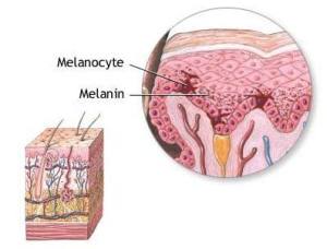 n cells against ultraviolet (UV) radiation by producing melanin. Melanin protects by absorbing harmful rays including UVA, UVB, UVC and blue light. Pigmentation of the skin are caused by accumulation of melanin-containing melanosomes in the basal layer of the epidermis. The number and activity of Human Epidermal melanocytes varies among individuals, influencing skin tone. Melanocytes are useful in studying melanoma, response to UV radiation, psoriasis and other skin diseases, skin trauma (e.g. wound repair, scars, burns etc.), cosmetic research (e.g. skin lightening compounds, skin protecting compounds).
Primary Epidermal Melanocytes:
Primary Epidermal Melanocytes are isolated from skin biopsies or cosmetic surgery procedures. The skin samples are enzymatically treated with trypsin or dispase to separate the epidermis from the dermis. The epidermis is further digested to release individual cells. The selective culture medium supports melanocyte growth while inhibiting the growth of keratinocytes and fibroblasts. Further, Melanocytes are purified by techniques like differential adhesion, flow cytometry or magnetic-activated cell sorting (MACS) based on specific surface markers.

iPSC-derived Melanocytes
Induced pluripotent stem cells (iPSCs) can be differentiated into melanocytes, providing an alternative source of these cells for research and therapeutic applications. Somatic cells (Fibroblasts, Peripheral Blood Mononuclear Cells etc.) are reprogrammed to pluripotent state by introducing OCT4, SOX2, KLF4 and CMYC. iPSCs are differentiated towards melanocytes using specific growth factors like Wnt3a, c-Kit, Endothelin-3 that promotes melanocyte development.
Characterization of Melanocytes:
Characterizing melanocytes involves several techniques to confirm their identity and functionality.
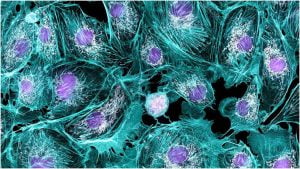
- Morphological Assessment: Melanocytes are fully differentiated and pigmented cells. Melanocytes exhibit dendritic morphology.
- Immunocytochemistry: Cells are stained for specific markers such as Melan-A, tyrosinase, HMB-45 (Human Melanoma Black-45), S100, MITF (Microphthalmia-associated Transcription Factor) to confirm melanocyte identity.
- Melanin Production: Synthesis of melanin is a key functional characteristic of Melanocytes. This is often assessed by using staining techniques e.g. Fontana Masson stain. The melanin reduces ammoniacal silver nitrate to metallic silver, resulting in black deposits.
- Gene Expression Analysis: Quantitative PCR or RNA sequencing are used to analyse the expression of melanocyte-specific genes e.g. Tyrosinase (TYR), tyrosinase related protein 1 (TYRP1), tyrosinase related protein 2 (DCT), premelanosomal protein (PMEL), glycoprotein NMB (GPNMB), and melanoma antigen 1 (MLANA)
- Flow Cytometry: This technique can be used to assess the expression of surface markers and quantify the purity of the melanocyte population. Melanocyte-specific markers e.g. Melan-A, tyrosinase, HMB-45, S100, MITF etc. are used for this quantification.
- Functional Assay: Alpha-Melanocyte Stimulating Hormone (α-MSH) bind to the MSH-receptors expressed by the melanocytes. Assessment of the ability of melanocytes to respond to melanogenic stimuli (e.g. α-MSH) and produce melanin.

Applications of Melanocytes
Melanocytes have diverse applications in skin biology research, medicine and cosmetics.
- Skin Disorders: Research on melanocytes helps in understanding pigment disorders like vitiligo, albinism, melasma, Addison’s disease, post-inflammatory pigmentations etc. This research is required to develop new treatments for these disorders.
- UV Protection Studies: Melanocytes play a crucial role to protect skin from UV radiations. Melanocytes are used to study the skin’s response to UV radiation and develop protective sunscreen against UV-induced damage.
- Cosmetic Testing: Cosmetic formulations to reduce hyperpigmentation target to inhibit melanin synthesis in melanocytes. By exposing these formulations to melanocytes in vitro, change in melanin synthesis give the effectiveness of the products. Melanocytes are also used to evaluate the efficacy of anti-aging formulations which aims to reduce age spots and skin-lightening.
- Cancer Research: Melanoma is a kind of skin cancer that initiates in melanocytes. Melanocytes are used to study mechanism of melanoma development and identifying novel therapies.
- Drug screening: Melanocytes are used in high-throughput screening assays to identify novel chemical entities that affect pigmentation.
Conclusion
Human epidermal melanocytes are invaluable tool in skin biology research, cosmetics and regenerative medicines. The significance of melanocytes to drive innovations in dermatology, regenerative medicine and therapeutic interventions for skin disorders and skin cancers is well-established. At Kosheeka, we committed to provide solutions for this ground-breaking research. We provide well-characterized, viable Melanocytes which are amenable to high-throughput drug screening. For more information about our customized cell culture services and to learn how we can support your research needs, please contact us or call us at +91-96543-21400
Frequently Asked Questions
Q.What are human primary melanocytes?
Human primary melanocytes are melanin-producing cells located in the basal layer of the epidermis. They produce melanin, the pigment that gives color to skin, hair, and eyes. Melanocytes play a crucial role in protecting skin cells against ultraviolet (UV) radiation by absorbing harmful rays including UVA, UVB, UVC, and blue light.
Q.How are primary epidermal melanocytes isolated?
Primary epidermal melanocytes are isolated from skin biopsies or cosmetic surgery procedures. The skin samples are enzymatically treated with trypsin or dispase to separate the epidermis from the dermis. The epidermis is further digested to release individual cells. Melanocytes are then purified using techniques like differential adhesion, flow cytometry, or magnetic-activated cell sorting (MACS) based on specific surface markers.
Q.What are iPSC-derived melanocytes?
Induced pluripotent stem cells (iPSCs) can be differentiated into melanocytes, providing an alternative source for research and therapeutic applications. Somatic cells are reprogrammed to a pluripotent state by introducing factors like OCT4, SOX2, KLF4, and CMYC. These iPSCs are then differentiated into melanocytes using specific growth factors like Wnt3a, c-Kit, and Endothelin-3.
Q.How are melanocytes characterized?
Melanocytes are characterized using several techniques to confirm their identity and functionality
- Morphological Assessment: Melanocytes exhibit dendritic morphology and are fully differentiated and pigmented.
- Immunocytochemistry: Cells are stained for specific markers such as Melan-A, tyrosinase, HMB-45, S100, and MITF.
- Melanin Production: Melanin synthesis is assessed using staining techniques like Fontana Masson stain.
- Gene Expression Analysis: Quantitative PCR or RNA sequencing analyzes the expression of melanocyte-specific genes.
- Flow Cytometry: This technique assesses the expression of surface markers and quantifies the purity of the melanocyte population.
- Functional Assay: Melanocytes’ ability to respond to melanogenic stimuli (e.g., α-MSH) and produce melanin is evaluated.
Q.What are the applications of melanocytes?
Melanocytes have diverse applications in skin biology research, medicine, and cosmetics:
- Skin Disorders: Research on melanocytes helps understand pigment disorders like vitiligo, albinism, melasma, and more, aiding in developing new treatments.
- UV Protection Studies: Melanocytes are used to study the skin’s response to UV radiation and develop protective sunscreens.
- Cosmetic Testing: Cosmetic formulations targeting melanin synthesis in melanocytes are tested for their efficacy in reducing hyperpigmentation and age spots.
- Regenerative Medicine: iPSC-derived melanocytes are used in therapies for pigmentation disorders and skin grafting.
- Cancer Research: Melanocytes are used to study melanoma, a skin cancer originating in these cells, to identify novel therapies.
- Drug Screening: Melanocytes are utilized in high-throughput screening assays to discover new compounds affecting pigmentation.
Q. Why are human epidermal melanocytes significant?
Human epidermal melanocytes are invaluable for advancing skin biology research, cosmetics, and regenerative medicine. Their role in protecting against UV radiation, understanding pigment disorders, developing sunscreens, and studying melanoma is crucial. Innovations in dermatology and therapeutic interventions for skin disorders and cancers heavily rely on melanocyte research.

