Kidney is a vital organ of the body; it acts as a filter to evacuate squander items from blood and produce urine. It regulates blood pressure and electrolytes in the body. There are more than 20 sorts of epithelial cells in kidney. Specialized immune cells, endothelial cells and interstitial cells in kidney play pivotal parts in kidney functions. The nephron, filtration unit of mammalian kidney, comprises of glomerulus.
Types of Kidney Cells:
Kidneys are composed of millions of nephrons, which contains diverse sorts of cells:
Glomerular Cells:
- Podocytes: Podocytes are specialized glomerular epithelial cells of kidney, plays a vital part in retaining plasma protein from entering in urinary filtration.
- Glomerular Endothelial Cells: These specialized cell lines the glomerular capillaries of kidney.
- Mesangial Cells: Mesangial cells line and support glomerular capillaries.
Tubular Epithelial Cells:
- Proximal Tubule Cells: Proximal tubule cells compose 60% of kidney, it lines the proximal tubule. Its pivotal for reabsorption of nutrients in kidney.
- Loop of Henle Cells: These cells are significant to retain sodium in the kidney.
- Distal Tubule Cells: Distal tubule cells are tall cuboidal epithelial cells, involved in controlling large particles in kidney.
- Collecting Tubule Cells: Collecting tubule carry urine from nephron to bladder and ureter. Collecting tubule cells are involved in acid-base homeostasis.

Human Kidney Epithelial Cell Lines:
- HK-2 (Human Kidney-2): HK2 cells are epithelial cells inferred from ordinary human male kidney. HK2 cells are immortalized by transduction with human papilloma infection 16 (HPV-16). HK2 cells are commonly utilized in renal toxicology research.
- HEK293 (Human Embryonic Kidney 293): HEK293 cell line display epithelial morphology, isolated from kidney of human embryo. HEK293 cell line is used in 3D cell culture, toxicology and transfection studies.
- Caki-1: Caki-1 cell line display epithelial morphology, derived from kidney tissue of male persistent with clear cell carcinoma. Caki-1 cell line is commonly utilized in renal cancer research, toxicology and 3D cell culture.
- RPTEC/TERT1 (Renal Proximal Tubule Epithelial Cells): RPTEC/TERT1 cells are derived from normal human renal proximal tubule of a male, hTERT-immortalized epithelial cells. These cells are utilized in 3D organoid (tubuloids) culture, renal physiology and toxicology.
- ACHN: ACHN cells are derived from metastatic tissue of a male renal adenocarcinoma patient. ACHN cells are utilized in renal adenocarcinoma studies and 3D cell culture.
- A-498: A-498 cells are epithelial cells derived from kidney tissue of a female renal cell carcinoma patient. A-498 cells are used in transfection studies.
- 786-O: 786-O cells are derived from kidney of a male patient with clear cell adenocarcinoma. 786-O cells are utilized to study renal carcinoma.
- HKC-8: HKC-8 cells are derived from human renal cortex cells and immortalized with SV40 T antigen. HKC-8 cells are utilized for nephrotoxicity studies.
- HRCE (Human Renal Cortical Epithelial Cells): HRCE cells are derived from human renal cortex. HRCE cells are utilized for nephrotoxicity, renal physiology and renal infection studies.
Primary Kidney Epithelial Cells:
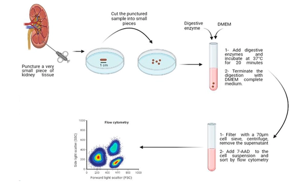
Primary kidney epithelial cells are isolated from fresh human kidney biopsies collected either from cadaver or surgical squander beneath sterile conditions. Kidney tissues are to begin with mechanically punctured, taken after by enzymatic treatment (collagenase, hyaluronidase, trypsin etc.). Primary kidney cells closely mimic the in vivo conditions, so are valuable in kidney disease modelling, nephrotoxicity, high throughput drug screening, kidney physiology and toxicology studies.

Figure 2: Schematic representation of isolation of viable cells from human kidney biopsy. Image Resource: PMID: 35620054
Human Induced Pluripotent Stem Cells (iPSCs)-derived Kidney Cells: Human iPSCs are differentiated towards kidney cells by using small molecules GSK3 inhibitor (CHIR99021), TTNPB, FGF9, EGF etc. iPSC-derived kidney cells are crucial for kidney disease modelling e.g. polycystic kidney syndrome, diabetic nephropathy etc; regenerative medicine, personalized medicine, and toxicology studies.

Characterization of Kidney Epithelial Cells: Kidney cells are characterized by distinctive strategies like morphological assessment, molecular characterization, functional assays etc.
Morphological Assessment: Kidney epithelial cells show apical-basal polarity, cuboidal morphology with distinct nucleus, which is assessed by phase-contrast microscope. Ultrastructure like microvilli, tight junctions, intracellular organelles of kidney epithelial cells are assessed by transmission electron microscope (TEM).
Molecular Characterization: Gene expression studies of kidney epithelial cells are assessed by RT-PCR or RNA sequencing. Kidney epithelial cells express specific markers such as E-cadherin, Aquaporin, N+/K+ATPase, cytokeratin like K7, K8, K18, and K19.

Protein Expression Characterization: Localization of kidney-specific proteins are assessed by immunocytochemistry against Na⁺/K⁺-ATPase on the basolateral layer or AQP2 on the apical layer etc. E-Cadherin, EpCAM, Aquaporin-1, integrin β1 and few intracellular cytokeratin markers are commonly assessed by flowcytometry.
Functional Characterization:
Sodium-Potassium ATPase Action: The action of Na⁺/K⁺-ATPase are measured by colorimetric or radioactive assay.
Tran epithelial Electrical Resistance (TEER): Integrity and permeability of epithelial monolayer are assessed by electrode to assess resistance across the cell layer.
Ion Flux Assay: The movement of particles like Na⁺, K⁺, or Cl⁻ across the epithelial cell layer are measured by utilizing ion-sensitive dyes.
Aquaporin Expression: The expression and function of aquaporin, are significant for water reabsorption in the kidneys, measured by utilizing fluorescence-based osmotic swelling assays.
Glucose Resorption: Uptake of glucose by kidney epithelial cells are measured by utilizing radioactive or fluorescently named glucose analogs (e.g., 2-deoxyglucose).
Protein Resorption: Protein resorption capability of kidney epithelial cells is measured by fluorescently tagged albumin uptake assay.
Applications of Kidney Epithelial Cells: Kidney epithelial cells are vital for renal physiology, drug metabolism, and disease modelling studies.
Drug Efficacy and Toxicity study: Kidney epithelial cells are utilized to assess nephrotoxicity, efficacy and safety of drugs.
Disease Modelling: iPSC-derived kidney cells are utilized to study kidney diseases like chronic kidney disease (CKD), Polycystic Kidney Disease (PKD), Aute Kidney Injury (AKI) etc.
Regenerative Medicine and Tissue Engineering: Kidney cells are utilized to replace damaged renal tissues in illnesses like CKD, AKI etc. 3D kidney organoids are utilized to study kidney diseases and embryonic development. Artificial Kidney transplantation is utilized to replace damaged kidneys in patients.
Drug Metabolism: Kidney epithelial cells are utilized to study about drug metabolism especially renal transporters and enzymes, which is significant for pharmacokinetics.
Biomarker Discovery: Kidney epithelial cells are utilized to study biomarkers identified in urine.
Conclusion:
Kidney epithelial cells are vital to study renal diseases, drug discovery and development. Kosheeka offers high-quality, 100% viable and functional kidney-epithelial cells. These cells are amiable to high throughput drug screening and 3D kidney organoid culture.
Frequently Asked Questions:
How Kosheeka support kidney research?
Answer: Kosheeka offers high-quality, 100% pure and functional kidney-epithelial cells, amenable to high-throughput drug screening and 3D kidney organoid culture.
What is Nephron?
Answer: Nephrons are functional unit of kidney.
What is drug-induced nephrotoxicity?
Answer: Few drugs cause harm to kidney, called as nephrotoxicity. In vitro kidney models are utilized to assess nephrotoxicity during drug discovery and development processes.
What are Podocytes?
Answer: Podocytes are specialized glomerular epithelial cells of kidney, plays a significant role in retaining plasma protein from entering urinary ultrafiltration.


