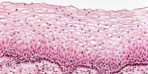
Endothelial cells fuse together to compose a monolayer named endothelium (Fig 1). This endothelium comprises the lining of the blood vessels and lymphatic vessels. Initially it was considered as epithelium. But the studies have established these cells as a different entity altogether due to their distinguishing features and functions from epithelial cells. It creates an interface between blood or lymph and the underlying tissue, thus governing the traffic across the tissue. But its role has been further delineated in regulating coagulation, maintaining vascular tone, promoting immune cell migration, controlling vascular permeability, and altering vessel diameter. Due to their involvement in cardiovascular diseases, endothelial cells are preferentially studied with respect to understanding the cardiovascular signaling mechanisms and developing their therapeutic options. But more applications of endothelial cells have been listed below.

Tumor Therapy
A major concern of tumor chemotherapy is the adverse effects on normal tissues due to the non-specific biodistribution of drugs.
The Enhanced Permeability and Retention (EPR) Effect: In the year 1986, two scientists- Matsumura and Hiroshi Maeda– discovered the accumulation of macromolecules in tumor tissue at high concentrations. They termed the phenomenon EPR. Further research delineated that tumor tissue vasculature is discontinuous or “leaky”. On the other hand, normal tissue vessels are more structured. Therefore, the leaky vasculature enhances permeability and more accumulation of molecules in a specific size range in the tumor tissue. This distinguishing feature allowed for the formulation of therapeutics in nanometer size to target the tumor tissue and eliminate the fatal off-target effects of the tumor therapy. Moreover, the lymphatic drainage is also defective in tumor tissue, thus prolonging the retention of chemotherapy drugs.
The Therapeutics Directed at Vessels: Owing to the role of tumor vasculature, scientists also prepared antibodies against VEGF and its receptor to disrupt the tumor vasculature, like Bevacizumab (anti-VEGF monoclonal antibody) and Pazopanib (anti-VEGF receptor tyrosine kinase inhibitor antibody).
Heterogeneity
Research has revealed that the endothelium assumes the structure and functions of the associated tissue. The endothelial cells express differential markers characteristic of the tissue. The heterogeneous nature of these cells extends further to the phenotypic variation. The endothelium displays different levels of permeability depending on the tissue. It is continuous with negligible permeability in the brain to confer protection. The layer becomes fenestrated in the gastrointestinal tract to enhance absorption. Endothelium is discontinuous with high permeability in the spleen. Such variability in structure and function is also evident in arteries, veins, arterioles, venules, and capillaries. Therefore, more researchers are diving into the study of the endothelial genome and proteome by single-cell sequencing. These studies are highly beneficial in basic understanding of tissue, vascular changes in pathologies, and the tissue/vessel-specific molecules for targeting therapeutics.
Inflammation
Immune cells or leukocytes migrate to the site of tissue injury or infection to destroy pathogens. But the leukocyte migration is absent in normal conditions owing to the role of endothelial cells in this process. In a pathological state, endothelium transitions from an anti-adhesive to an adhesive surface. They express adhesion molecules that interact with their corresponding ligands on the leukocytes. Thus, the endothelium aids in the migration process via the following steps.
Capture: Endothelium expresses selectin molecules, specifically E-selectin and P-selectin, that form weak interactions between endothelium and leukocytes. The purpose of this step is to get the leukocytes in the proximity of the endothelium.
Adhesion: The first step initiates a signaling cascade in the endothelium, upregulating adhesion molecules ICAM1 and VCAM1. These molecules mediate a firm adhesion of leukocytes to the endothelial layer.
Diapedesis: This step witnesses the movement of leukocytes over the endothelial layer by the continuous breaking and formation of bonds.
Transmigration: The leukocyte movement stops as they reach the junction between two endothelial cells. The junction is maintained by CD99, VE-cadherin, and JAM. Transient removal of these molecules occurs during the transmigration step. It loosens the junctions and increases the permeability to transport leukocytes to the underlying tissue.
Each step is vital for adequate immune responses. But in certain pathologies such as atherosclerosis, this mechanism turns dysfunctional, resulting in chronic inflammation.
Blood-Brain Barrier
The brain vasculature aims to restrict passage across the brain for neuroprotection and prevent neurotoxicity. Thus, the endothelial cells are non-fenestrated and form a continuous layer. The tight junction between two endothelial cells is selective to inhibit the passive diffusion. Combined together, the endothelium forms a near impermeable layer in the brain. This blood-brain barrier allows the transport of small molecular weight lipophilic molecules. Few biomolecules like glucose employ endothelial transporters, whereas large molecules use receptor-mediated endocytosis. Scientists have a special interest in this due to its implications in drug delivery. Studies have established that expression of ABC transporters reduces in certain neurological disorders such as Alzheimer’s disease and Parkinson’s disease. Dysregulation of this barrier also leads to chronic inflammation, resulting in multiple sclerosis. Such myriad roles of the endothelial layer have urged the researchers to study the transport pathways across the endothelium.
Coagulation
Endothelium also maintains vascular tone and hemostasis. Circulating platelets cannot adhere to the endothelial surface owing to its anticoagulant nature. Endothelial cells release nitric oxide and prostaglandin I2 that inhibit calcium ion release via the formation of cGMP and cAMP, respectively. Moreover, these cells secrete tissue factor pathway inhibitor and antithrombin III that prevent activation of coagulation factors. Endothelial injury exposes the matrix collagen for platelet binding. Additionally, endothelial cells also release vWF and platelet activating factor to promote platelet aggregation and activation, respectively. The endothelial matrix also contains tissue factor, which is also exposed during injury. Tissue factor binds to coagulation factors and initiates the coagulation cascade. These mechanisms switch the endothelium between anticoagulant and procoagulant surface. Any dysfunction in these processes leads to serious disorders such as atherothrombosis, stroke, occlusive diseases, etc. Hence, researchers are extensively studying the endothelial surface characteristics in a pathological state.
Angiogenesis and Vasculogenesis
Vasculogenesis and angiogenesis are the complex and dynamic processes of blood vessel formation (Fig 2).
Vasculogenesis: The process involves the formation of new blood vessels that occurs during the embryonic stage.
Angiogenesis: Angiogenesis follows the formation of blood vessels from the already existing vessels. The process could be categorized into sprouting angiogenesis and intussusceptive angiogenesis. Sprouting angiogenesis entails the formation of new branches of vessels from the already present vessels. Intussusceptive angiogenesis involves dividing a single vessel into two vessels.
Product-Related Queries, Or Partnership Inquiries
Angiogenesis is an essential process for growth and repair. Its dysfunction can cause ischemic disorders, malignant diseases, neurodegenerative diseases, and pathological wound healing.

(Image Source: https://www.ncbi.nlm.nih.gov/books/NBK53238/)
Conclusion
The endothelial cells are implicated in several disorders such as peripheral arterial disease, ischemic limb injury, cardiovascular diseases, strokes, diabetes, renal failure, etc. It necessitates research into their structure and function. The research on endothelium can generate new insights into tissue-specific pathways and their applications in the formulation of new therapeutics. In 2024, US FDA even granted approval to Humacyte SYMVESS, a tissue-engineered vessel, for the treatment of arterial injury and prevention of loss of limb. At Kosheeka, we provide primary endothelial cells at low passage to advance your endothelial cell research. Our team thoroughly characterizes and robustly evaluates the endothelial cells to ensure highly viable and functional cells. We also offer customer support and necessary guidance for culture and maintenance of these cells in culture to accelerate your research.
FAQ
What are the sources of endothelial cells?
Endothelial cells can be extracted from blood vessels. These blood vessels can belong to any tissue, such as glomerular microvascular cells, dermal microvascular cells, pulmonary endothelial cells, etc. However, HUVECs are considered the ideal model for in vitro study.
What are the functions of endothelial cells?
Endothelial cells act as a selective barrier between blood and the underlying tissue. Moreover, these cells also maintain permeability, control vascular tone, and change vessel diameter.
Where to procure endothelial cells?
Kosheeka provides primary endothelial cells isolated by a team of highly qualified and experienced personnel. The cells undergo rigorous testing and thorough characterization to deliver a quality-assured product.
