

Introduction
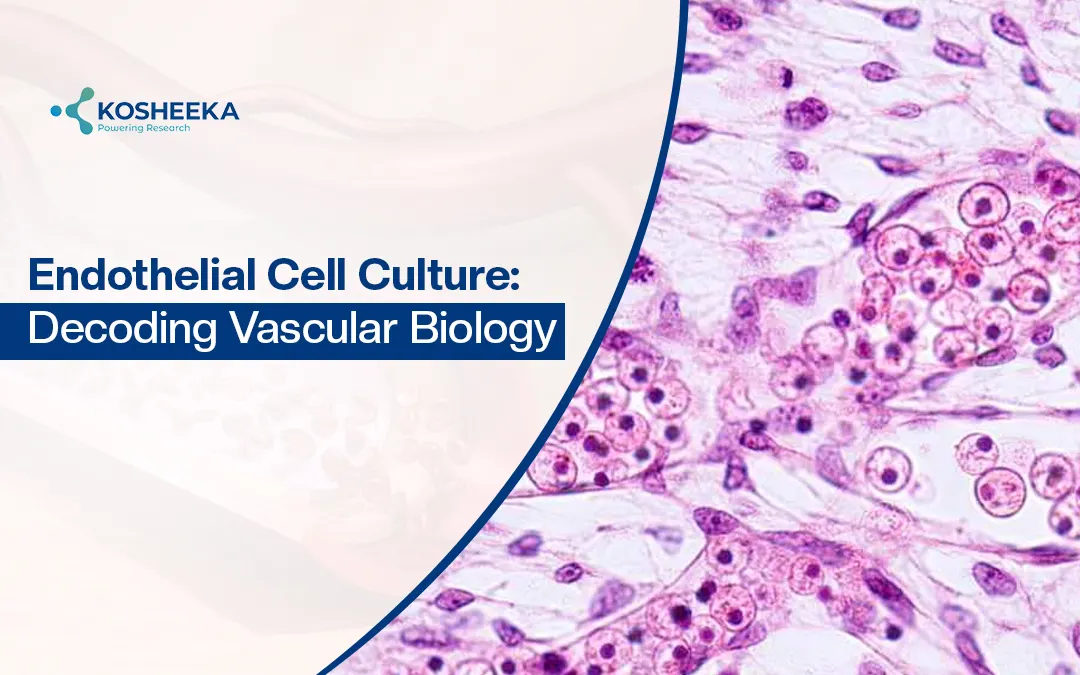
Endothelial cells form ‘tunica intima’- a monolayer of flat cells that forms the inner lining of the blood vessels, heart, and lymphatic vessels. The cells form a smooth endothelium surface that acts as a selective filter. Functionally, the layer maintains tissue homeostasis and facilitates the exchange of fluid, gases, immune cells, or other biological molecules.
In vascular biology, their function overcomes the functional barriers. The cells are widely distributed in endocrine organs and have a dynamic role in maintaining vascular tone and blood flow. In-vitro Endothelial Cell Culture is a critical technique for researchers to conduct cardiovascular, inflammatory, angiogenesis, and regenerative research.
FACT CHECK: Human endothelium collectively weighs similar to the liver. It forms the largest endocrine interface.
Understanding Endothelial Cells and Their Biological Functions
Endothelial cells form a single endothelium layer that line blood vessels and lymphatic vessels. The endothelium layer forms a surface for blood and tissue interaction. The location includes:
- Veins
- Arteries
- Capillaries
- Lymph capillaries
The two common terms, vascular endothelium and lymphatic endothelium, come up in vascular research. Hereby:
- Vascular Endothelium: Endothelial cells that form the blood vessel lining, help in blood flow
- Lymphatic Endothelium: Endothelial cells form the lymphatic vessel linings, which help lymphatic fluid flow
*Both have a crucial role in cardiovascular health.
What are the Key Physiological Functions?
Endothelial cell function depends on their location in the body. The key functions include:
Vasoconstriction & Vasodialation
- Endothelial cells aid in blood vessel relaxation
- Allows adequate blood flow in different body parts
- Adapt to internal (pressure against vessel walls) and external stimuli (temperature)
Influence Fluid Exchange
- Endothelium regulates permeability, allowing fluid movement between cells and tissues
- Enable an adequate barrier and protect the blood against toxins or other foreign particles
Thrombosis Prevention
- Thrombosis is characterized by blood clot formation that blocks blood flow
- Produces nitric oxide and prostacyclins, regulates vascular tone
- Maintains blood fluidity, prevents unnecessary blood clotting
Angiogenesis
- Endothelial cells are involved in new blood vessel formation
- Essential in processes like wound healing
- Regulates key factors like vascular endothelial growth factors (VEGFs), angiopoietins, fibroblast growth factors (FGFs)
FACT CHECK: Endothelial cells from distinct organs express distinct signature genes, which also act as biomarkers
What is Endothelial Dysfunction?
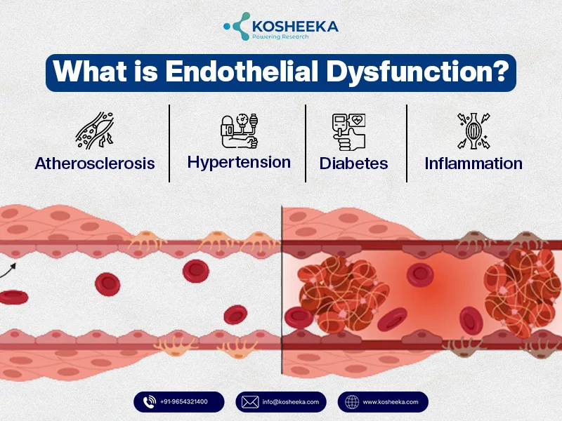
Endothelial dysfunction can be the earliest sign of vascular disease. It is characterized by nitric oxide bioavailability reduction, an increase in oxidative stress, pro-inflammatory cytokines, and a pro-thrombotic state. Various endothelial dysfunction-associated diseases include:
- Atherosclerosis: Chronic endothelial dysfunction and injury. High levels of LDL (bad cholesterol) lead to damage. The immune cells kick monocytes to the damaged parts of the endothelium. This forms monocyte clusters at the damaged spot that trigger inflammation. It forms plaque formation. Gradually, the plaque grows and narrows the artery. It raises the risk of blood clots
- Hypertension: Elevated blood pressure creates tension on the endothelium. Endothelial dysfunction and hypertension lead to NO impairment and increased oxidative stress. It leads to vasoconstriction and vascular remodeling.
- Diabetes: Hyperglycemia and insulin resistance lead to diabetes mellitus. Leads to an elevated level of Advanced Glycation End Products (AGEs) and ROS
- Inflammation: Endothelium dysfunction triggers immune response and activation of pro-inflammatory cytokines (TNF-α, IL-1β). It exacerbates vascular damage via triggering the expression of adhesion molecules or pro-thrombotic factors [1].
FACT CHECK: Endothelial dysfunction develops long before the appearance of plaque formation in cardiovascular disease.
What is the Importance of Endothelial Cell Culture?
Endothelial cell culture acts as an accurate research model for vascular diseases. Human primary endothelial cells (HPECs) are isolated from primary sources. For researchers, it acts as a useful tool for vascular studies, angiogenesis, wound healing, and high-throughput applications.
Why Primary Endothelial Cell Isolation Matters?
- Primary Endothelial Cell Isolation shares physiological resemblance with native tissue
- It enables researchers to develop an endothelial dysfunction disease model
- Unlike immortalized cells, primary endothelial cells have genetic, functional, and phenotypic characteristics
- Enable investigation of vascular tone and barrier function
- Development of pre-clinical data, before moving towards clinical application
What are the Common Sources for Endothelial Cell Isolation?
- Endothelial cells from the coronary artery and the aorta
- Umbilical vein endothelial cells (HUVECs)
- Microvascular endothelial cells
- Tissue-specific vascular isolation approaches
Kosheeka, India is a leading cell culture laboratory that isolates and supplies primary and secondary cells for research purposes. The laboratory is GMP certified and maintains aseptic conditions and controlled environment. The cells undergo stringent quality checks before dispatch.
How are Endothelial Cells Isolated for Vascular Studies?
Endothelial cell isolation involves a multi-step process. The steps include:
Tissue Harvesting
- Tissue excision from the heart
- Tissue washed with ice-cold PBS (blood removal)
- Tissue is dissected to remove excess fat or unwanted adherent tissue
- In sterile conditions, tissues are minced
Enzymatic Digestion
- Collagenase treatment, minced tissue incubated in 0.5-1mg/ml collagenase for 45-60 min
- Cells passed through a strainer (70-100µM), removing undigested chunks
- Cells were incubated in 0.25% trypsin-EDTA to obtain a single cell suspension
Immunomagnetic Separation
- All cell suspensions are incubated with immunoglobulins at 4°C
- Blocks non-specific receptors
- Cells incubated with endothelial-specific markers (CD31, CD105, Isolectin B4)
- Attach the cells in microbeads. Separate with the help of a magnetic column
Culture
- Cells are added to a pre-coated tissue culture flask with complete medium
- Endothelial growth medium is used with FBS
- Incubated at 37°C, in presence of 5% CO2, and adequate humidity
What are the Major Challenges in Endothelial Cell Isolation?
The major limitations include:
- Phenotypic drift during culture
- Variability of cell features based on donor variability
- Maintenance of endothelial cells over passages is complex
What is the Application of Endothelial Cell Culture?
Endothelial cell culture has a wide range of applications. This includes:
Establishing 2D Endothelial Cell Culture
- Involves developing a monolayer culture
- Various research applications like disease modeling, drug screening, targeted therapy, regenerative medicine, etc.
Developing 3D Vascular Models/ Organ-on-Chip
- Mimics blood flow & shear stress
- Establishing a co-culture system with fibroblast or smooth muscle cells
- Developing a microfluidic vascular platform
Understanding Endothelial Cell Migration
- Endothelial cell migration is crucial in wound healing study
- Unveiling chematactic signaling pathway
- Angiogenesis and developing VEGF-mediated cellular migration
Drug Development
- Drug screening, toxicology testing, and development of targeted therapy
- Understanding endothelial cell proliferation, the role of the cell cycle in vascular repair
- Developing an anti-inflammatory vascular drug
- Understand the key differentiation between regeneration (regenerative medicine) and pathological conditions (cancer-associated angiogenesis)
What are the Common Research Assays?
Endothelial cells use in in-vitro assays includes:
- Scratch/ Wound Healing assay
- Tube formation assay
- Transwell migration studies
- Cytotoxicity Study
- Tumor angiogenesis
- Diabetic wound healing
- Vascular graft development
- Regenerative research
Conclusion
Endothelial cell culture has a pivotal role in vascular biology and translational medicine. The in-vitro culture enables researchers to have a better understanding of vascular science. Various aspects like Endothelial Dysfunction, inflammation, tissue regeneration, oncology, systemic disease progression, etc., are better understood. It enables the development of advanced diagnostic markers, drug development, targeted therapy, and regenerative medicine.
References
- Ray A, Maharana KC, Meenakshi S, Singh S. Endothelial dysfunction and its relation in different disorders: Recent update. Health Sciences Review. 2023 Jun 1;7:100084.
FAQ’s
Q- How do Endothelial Cells Help in Vascular Research?
Endothelial cells form the inner lining of the blood vessels. They aid in regulating blood vessel function, vascular permeability, angiogenesis, and inflammation. These cells are core responders to inflammatory and metabolic changes. It enables a detailed understanding of cardiovascular disease, endothelial dysfunction, tissue repair, etc.
Q- What is Endothelial Dysfunction?
Endothelial dysfunction are characterized by impaired vascular endothelium functioning. This leads to reduced nitric oxide production and increased inflammation. Endothelial dysfunction can be an early sign of atherosclerosis, diabetes, stroke, and hypertension development.
Q- Why Do Researchers Prefer Primary Endothelial Cell Isolation Over Immortalized Cell Lines?
Primary endothelial cells share better physiological and genetic relevance with native tissue biology. These cells enable better understanding and predictability related to endothelial cell migration, proliferation, disease modeling, and therapeutics. Immortalized cells are useful in developing large-scale experimentation. They are subjected to phenotypic drift.
Q- Is Endothelial Cell Culture Used in Regenerative Research?
Endothelial cell culture models help researchers to understand tissue regeneration, vascular repair, anti-inflammation, angiogenesis, and wound healing. The development of advanced 3D culture systems and organ-on-chip technologies has enabled new possibilities in developing vascularized tissue engineering approaches.



Endothelial cell culture models are becoming increasingly important for understanding how vascular dysfunction contributes to conditions like inflammation and cardiovascular disease. I also think it’s interesting how advancements in 3D cell culture and tissue engineering are making these models more physiologically relevant compared to traditional 2D systems. This kind of research could really improve the reliability of preclinical studies in the future.
Yes absolutely correct. For any cell culture needs, reach out:https://www.kosheeka.com/