
Human dermal fibroblasts (HDFs) have a central role in extracellular matrix (ECM) production, wound healing, tissue remodeling, and maintaining skin homeostasis. In in-vitro research, human dermal fibroblast cells are established models for exploring skin aging, fibrosis, drug discovery, regenerative medicine, and tissue engineering. Depending on the research requirements, dermal primary cells or cell lines are used. Careful consideration of donor variability, cell passage number, cell line, culture condition, etc., is essential to gain a desirable research outcome. The current article highlights essential insights, characteristics of the cells, applications, and best practices in the laboratory using HDFs.
What are Human Dermal Fibroblasts?
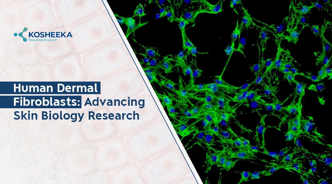
Human dermal fibroblasts are primary cells isolated from the human skin dermal layers. These cells are spindle-shaped and exhibit adherent culture properties. In in-vitro culture condition, the cells form a confluent monolayer with overlapping growth patterns on reaching confluency. Functionally, these cells have a vital role in the synthesis of ECM components, including collagen and elastin, mediate wound healing by fibronectin-mediated migration, and interact with epidermal cells during tissue repair and hair follicle development.
These cells demonstrate mesenchymal-like properties and express similar surface markers like CD73, CD90, and CD105. Other fibroblast-specific markers include vimentin, fibronectin, and collagen type 1. Researchers widely use these cells for investigating dermal homeostasis, degenerative skin conditions, pathological scarring, etc.
HDFs vs HDFs Cell Line
Selection between Human Dermal Fibroblast cell lines and HDFs is crucial for the appropriate designing of in-vitro research. Although both models share fibroblast-specific characteristics, they considerably differ in their origin, proliferative capability, physiological relevance, and experimental application.
Primary Fibroblasts vs Human Fibroblast Cell Line: Key Differences
Features | Primary Fibroblasts | Fibroblast Cell Line |
| Source | Isolated directly from human dermal layer | Primary cells transformed and immortalized into cell line |
| Physiological Relevance | Closely mimics physiological relevance with origin | Might exhibit altered biological function |
| Proliferative Capacity | Limited | Infinite, can divide into multiple passage |
| Donor | High, feature changes based on donor age, genetic variability, health condition | Low, provide high experimental consistency |
| Reproducibility | Moderate due to biological variation | High across the experiments or laboratories |
| Maintenance | High maintenance | Low in comparison with primary culture |
| Application | Skin biology, wound healing, fibrosis, regenerative medicine, disease modeling | Drug screening, toxicity testing, molecular studies, assay development, and routine laboratory research |
Which Model Should Researchers Choose?
The choice between primary fibroblast and Human Dermal Fibroblast Cell Lines depends on the research requirement and objective. Primary cells are a relevant model when the experiment is in a translational state for clinical studies. It is suited for experimentation such as skin regeneration, disease modeling or mechanism of aging. The dermal cell line is suitable where there is a need for high reproducibility, large sample size, prolonged culture, biomolecule production or high-throughput screening.
In advanced research, scientists combine both models for experimentation to strengthen biological findings.
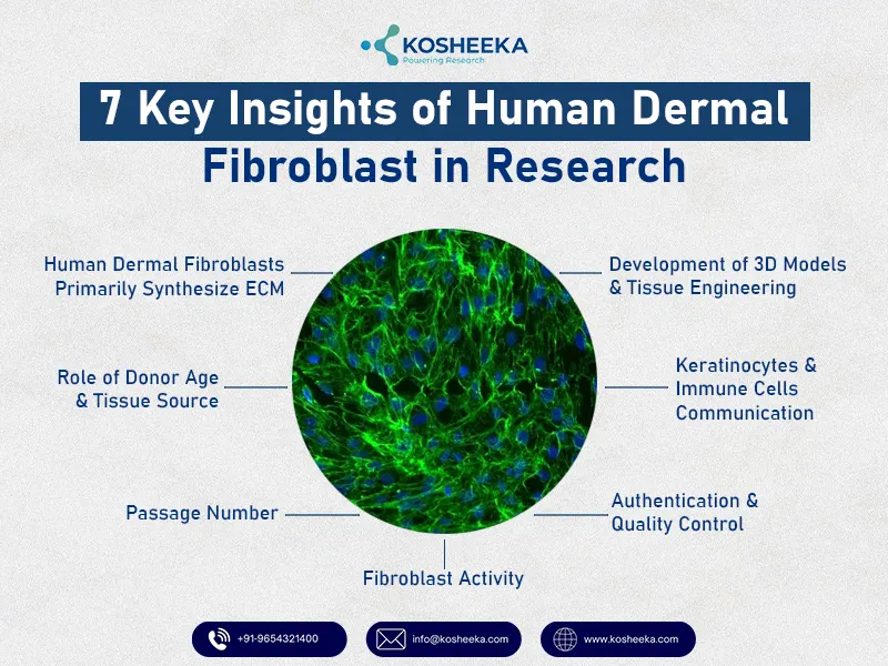
7 Key Insights of HDFs in Research
In biomedical and regenerative research, Human Dermal Fibroblast cells play a pivotal role. These cells are primarily responsible for maintaining skin structure, regulating tissue repair, ECM remodelling, regulating inflammation, and promoting cellular communication. The key insights include:
Human Dermal Fibroblasts Primarily Synthesize ECM
HDFs synthesize and organize ECM key components, including collagen type 1 and III, fibronectin, elastin, laminins, and glycosaminoglycans. They are responsible for maintaining intact skin, its elasticity, and resilience. ECM regulates multiple cellular signalling pathways, cell adhesion, migration, proliferation, and differentiation.
Role of Donor Age and Tissue Source
The researcher must take into account the donor age and the tissue source. Every fibroblast does not behave identically. The age, anatomical location, health status, and genetics influence its characteristics. For instance, neonatal fibroblasts exhibit rapid proliferation and collagen synthesis and possess greater regenerative capacity. At the same time, aged fibroblasts display reduced proliferative potential.
Passage Number
Prolonged culture, that is, culture with higher passage numbers often exhibits phenotypic, genotypic, or functional variations. In case of a higher passage number, slowed proliferation, altered morphology, or reduced production of collagen occurs. Similarly, reduced responsiveness to growth factors or increased expression of senescence-associated biomarkers is exhibited.
Fibroblast Activity
In biomedical and regenerative research, fibroblasts have an intrinsic role in wound healing and scar formation. Fibroblast secretes various cytokines, chemokines and various growth factors like FGF, VEGF. These bioactive molecules actively take part in inflammation, tissue remodeling or angiogenesis. While normal fibroblast cells have crucial mechanistic functions, their dysfunction can cause diseased conditions. Some examples include fibrosis, keloids, or hypertrophic scars.
Development of 3D Models and Tissue Engineering
The cells are widely used for the development of 3D skin constructs that can replicate skin architecture and cellular interaction. Fibroblasts can establish dermal compartments for producing ECM proteins and supporting a structural scaffold. Used in regenerative medicine research (cosmetic testing, disease modeling, wound healing, biomaterial evaluation, and pharmaceutical research).
Keratinocytes and Immune Cells Communication
HDF, by the secretion of various active bioactive components, facilitates communication with neighbouring keratinocytes, melanocytes, endothelial cells, and immune cells. The cells facilitate cross-talk to maintain epidermal homeostasis, angiogenesis, inflammatory response, tissue regeneration and pigmentation. Disruptive communication leads to chronic wounds, fibrosis, inflammatory diseases and impaired tissue regeneration.
Authentication and Quality Control
To gain reproducible and reliable experimental outcome. Misidentified data, genetic drift, mutations, or microbial contamination alter data integrity and reproducibility. Fibroblast characterization is a crucial element. Common fibroblast biomarkers include CD44, CD73, CD90, CD105, and vimentin. Regular mycoplasma testing, maintaining a sterile environment and maintaining a standard cryopreservation protocol. Further, adequate documentation is crucial, including donor information, culture conditions, culture media, and passage number. This supports reproducibility and transparency of the data.
*Kosheeka, India, is one of the leading animal cell culture laboratories that isolates and supplies primary fibroblast cells for research use. The cells are isolated and maintained under strict laboratory guidelines and undergo stringent quality checks.
Human Dermal Fibroblast Culture Protocol: Best Laboratory Practices
The Standards of the Human Dermal Fibroblasts Culture Protocol Involve:
- HDFs-specific medium such as DMEM supplemented with 10% FBS and 1% pen-strep antibiotic
- Incubation in CO2 incubator- 5% CO2, 37°C, and adequate humidity
- Seeding cells with adequate density ensures uniform attachment and avoids over-confluency
- Regular cell morphology monitoring. Fibroblast cells appear as spindled-shaped
- Routine change of medium to maintain adequate nutrient supply
- The cell is passaged with 80-85% confluency
- Experimental design with a lower passage number of the cells
- Strictly maintain sterile conditions, controlled environmental conditions, and maintain a contamination-free culture
- Maintain a regular record including donor information, passage number, media composition, seeding density, and culture duration
Conclusion
HDF is a versatile model for researchers in biomedical and regenerative research. The cells have a diverse range of applications, ranging from maintaining normal skin integrity to treating diseased conditions. Despite the easy availability of primary fibroblast cells, maintaining them in vitro is quite complex. Researchers have elaborated on the use of fibroblast cell lines for availability, convenience, ease of maintenance in vitro, and suitability in designing large-scale studies and data reproducibility.
FAQ’s
Q- What is the Function of HDFs?
HDFs are the primary cells of the skin’s dermis. Its main function is the synthesis of ECM components, including collagen types I and III, fibroblasts, elastin, and fibronectin. These cells maintain intact skin, its elasticity and defense mechanism.
Q- What Culture Medium is Commonly used for HDFs?
Dulbecco’s Modified Eagle Medium (DMEM) + 10% fetal bovine serum + 1% penicillin-streptomycin. The culture medium choice might differ based on specific experimental needs.
Q- Why does Passage Number Matter in In-Vitro Culture?
Passage numbers have a crucial role in experimental outcome accuracy. With lower passage numbers, phenotypic and genetic stability is inevitable. This ensures experimental outcome reliability. With an increase in passage number, fibroblasts might undergo cellular senescence, transcriptomic changes, morphological alterations, and genetic drift.