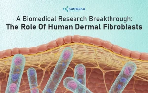
Introduction
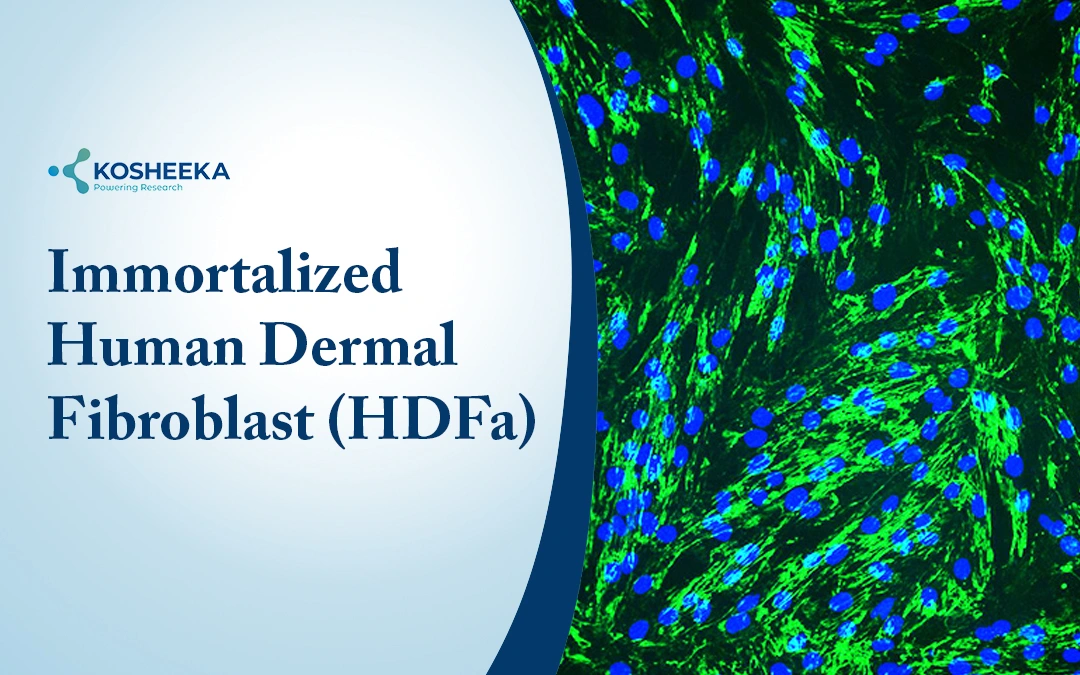
The advances in dermatological research primarily rely on human dermal fibroblasts as a powerful cellular model to study skin physiology, aging, and wound healing.
Human dermal fibroblast (HDFa) has active involvement in the production of extracellular matrix (ECM). They have a primary role in connective tissue networking in skin. The application of Human Dermal Fibroblast (HDFa) in primary and secondary animal cell culture research has enabled researchers to unveil the underlying science behind skin biology. It involves understanding physiological conditions, disease mechanisms, and therapeutic responses. In fact, in the past few decades, regenerative medicine knowledge has significantly evolved with dermal fibroblast research.
Structure and Biological Role of Human Dermal Fibroblasts
HDFa cells are an indispensable element in skin biology research. They are considered the gold standard model for understanding skin disease, aging, wound healing, toxicology, cosmetic development, and regenerative research. With research progression, the need for a more complex in-vitro system is required. Immortalized HDFa creates a stable and renewal source of fibroblast suitable for research applications.
The structure and characteristics include:
- Morphology: Spindle-shaped, elongated cells with prominent cytoplasm, active rough endoplasmic reticulum (ER), and Golgi apparatus for protein synthesis
- Location: Located in the dermis layer of the skin
- Characterization: Biomarker for identification is vimentin
- Cell Types: HDFa have cellular heterogeneity, i.e., they include upper papillary fibroblast and lower reticular fibroblast. The distinct subtypes have different mechanisms in skin tissue homeostasis.
- Function: Major function ECM synthesis, collagen and elastin production, tissue integrity and skin homeostasis
Human Dermal Fibroblast Cells Types and Culture Characteristics
Based on cell source and type, HDFa are subcategorized as primary cells and immortalized (secondary) cell lines. Both cell types have distinct characteristics and culture conditions. This includes:
Primary HDFa
- Isolated from dermal tissue sample (skin)
- Mimics native cellular morphology and physiological conditions that mimics humans skin
- Closely represents an in-vivo microenvironment, suitable for natural skin biology study
- Exhibits limited proliferative properties, limited life span
- Represents normal gene expression patterns and ECM production
- More vulnerable to the surrounding conditions (environmental and cultural medium)
- More prone to contamination (bacterial, viral, mycoplasma
Secondary HDFa
- Human Dermal Fibroblast Cell Lines are obtained from primary HDFa immortalized with hTERT and CDK4R24C viral infection
- Modified cells pooled for cell selection and expansion
- Immortalized cells are capable of continuous proliferation, which allows long-term experimental planning
- High experimental reproducibility and consistency in the results
- Easier to maintain in controlled laboratory conditions in comparison with primary culture
- Wide range of applications for obtaining preclinical study data. E.g., drug screening, toxicology, high-throughput screening, molecular pathway studies
- Might exhibit altered gene expression, cellular response and metabolic activities in comparison with primary fibroblast
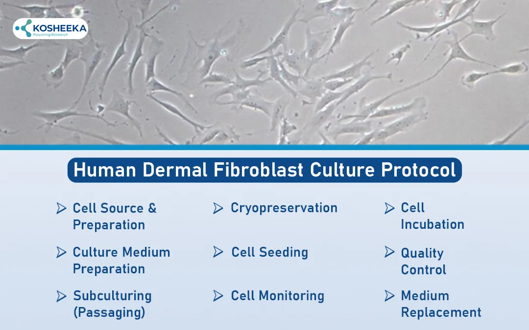
Human Dermal Fibroblast Culture Protocol
The isolation of human dermal fibroblast culture involves a multi-step process. This includes:
- Cell Source and Preparation: Cells obtained from a certified cell repository or isolated from a human dermal tissue sample. Cryopreserved cells vials are quickly thaw at a 37 °C water bath for 1–2 minutes
- Culture Medium Preparation: The complete culture medium is prepared, which includes medium +10% FBS and 1% antibiotic. The medium used includes DMEM or fibroblast basal medium.
- Cell Seeding: The thawed cell suspension is resuspended in a pre-warmed medium and centrifuged. Cell seeding in an appropriate culture flask/plate.
- Cell Incubation: Cells incubated in a humidified CO₂ incubator at 37 °C in presence of 5% CO₂
- Cell Monitoring: The cells are observed using a phase-contrast microscope for determining morphological relevance and adherence. The spindle-shaped morphology reflects the appropriate morphological relevance
- Medium Change: Culture medium is replaced at an interval of 2-3 days (removes metabolites and waste)
- Subculturing (Passaging): The cells, upon reaching 80-85% confluency, are passaged. Adherent cells detached using trypsin-EDTA solution for 2-3 min at 37 °C. Fresh medium added to neutralize trypsin effect. Cell reseeding is done in a new flask, cells are suitable for using experimental condition
- Cryopreservation: For long-term storage, cells were resuspended in freezing medium including serum and cryoprotectant. Cells stored in liquid nitrogen
- Quality Control: The cells were thoroughly checked for morphological accuracy, growth rate, and presence of contamination (bacteria, fungus, or mycoplasma) [1]
Research Applications of Human Dermal Fibroblasts
The research application of the Human Fibroblast Cell Line includes:
- Wound healing and tissue repair mechanisms
- Activation and reprogramming of fibroblasts into platelets
- Identification of surface biomarkers, differentiation between chondrocytes and synovial cells
- Skin aging, anti-aging therapeutics, and regenerative research
- Develop protocols for tissue engineering, synthetic collagens, and biomaterials.
- Understanding molecular mechanisms in gene regulation, activation, and epigenetic modification. microRNAs expression or histone ubiquitination
- Drug screening and toxicological studies
- Oncology and immunological studies
- Insights into disease progression and underlying cause, e.g., UVA effects, environmental stressors, premature cell senescence, tumour cell pluripotency markers, etc.
Role of Human Dermal Fibroblasts in Regenerative Medicine
Skin Regeneration Research
- Skin regeneration and repair of existing cells
- Active production of ECM components: collagen, elastin, and fibronectin
- Skin structure and elasticity
- Regenerative mechanisms in studies: Wound healing, tissue remodeling, and scar formation
- Regulates cell signaling and interacts with other skin cells (keratinocytes), skin regeneration
Tissue Engineering and Scaffold Development
- Human dermal fibroblasts used in tissue engineering
- Development of artificial or bioengineered skin substitutes
- Seeding of Fibroblasts into biocompatible scaffolds that mimic the ECM
- Scaffolds provide structural support, allow fibroblasts to proliferate and produce matrix proteins
- Research application in burn treatment, chronic wound management, and reconstructive surgery.
Cell-Based Therapeutic Approaches
- Dermal fibroblasts for cell-based therapies restore damaged skin tissue
- Fibroblast transplantation and fibroblast-derived bioactive factors stimulate tissue repair, angiogenesis, and extracellular matrix regeneration.
- Potential use in personalized regenerative therapies and advanced biomedical research
It is crucial to maintain and use superior quality cells for research use. Kosheeka, India, is among the renowned research laboratories that maintain and supply various primary and secondary cells for research purposes.
Advantages and Limitations of Using Human Fibroblast Cell Lines
Advantages
- Ease in culture medium
- High reproducibility rate
- Long-term proliferation
- Widely used experimental model
- Cost-effective in comparison with the in vivo model
Disadvantages
- Altered cellular characteristics
- Limited physical relevance
- Loss of functional properties with passage
- Lack of complex tissue interaction
- Genetic drift
Future Perspectives in Skin Biology Research
- 3D skin models
- Organoid model
- Development of advanced cell culture systems
- Regenerative application (Stem cell therapy or Exosome Therapy)
Conclusion
HDFa is derived from human skin’s dermis. It is critical in producing ECM, elastin and collagen synthesis, and fibroblast networking. In animal cell culture research, human dermal fibroblast cells play a crucial role in skin biology research. The dermal cell lines aid in studying various aspects, including dermatology, toxicology, cosmetics, and regenerative mechanisms.
References
- Kisiel MA, Klar AS. Isolation and culture of human dermal fibroblasts. InSkin tissue engineering: methods and protocols 2019 May 31 (pp. 71-78). New York, NY: Springer New York.
FAQ’s
Q- How do Human Dermal Fibroblast Cells AID in Skin Biology Research?
Human dermal fibroblast cells are connective tissue cells that are responsible for ECM formation. The cells promote collagen and elastin synthesis. The cells serve as an essential source for skin biology research, such as wound healing, tissue regeneration and repair mechanisms.
Q- What are the Research Applications of Dermal Fibroblast Cells?
The major research application includes skin aging, wound healing mechanisms, skin regeneration, repair mechanisms, skin homeostasis, ECM remodelling, drug testing, and toxicology profiling.
Q- How are Dermal Fibroblast Cells Important in Regenerative Medicine?
Dermal fibroblast cells in regenerative medicine help in various aspects. This includes EXM components, supporting skin regeneration, and tissue repair mechanisms. The cells are useful in obtaining preclinical data related to tissue engineering, cell-based therapies, and artificial skin development.


The use of dermal fibroblasts in tissue engineering is fascinating, especially considering their role in creating 3D models for drug testing. I think these models could revolutionize the way we approach disease research.
Absolutely—dermal fibroblasts are key players in building realistic 3D tissue models. Their ability to mimic native skin architecture makes drug testing more predictive and could significantly advance disease research and personalized therapies.
I’ve been absent for some time, but now I remember why I used to love this site. Thank you, I’ll try and check back more frequently. How frequently you update your web site?
Thank you so much for coming back—we’re really glad to have you here again! 😊
We update our website every week, sharing fresh and interesting topics, insights, and the latest developments in the field. There’s always something new to explore, so definitely check back regularly—we’d love to have you with us more often!
I’ve recently started a web site, the information you provide on this site has helped me tremendously. Thanks for all of your time & work.
Glad to hear from you. We believe in maintaining authenticity of the information. For more information Visit:https://www.kosheeka.com/