Introduction
The human body relies on specialized cellular function, well-coordinated remarkably at the cellular and molecular levels. At first glance, epithelial and endothelial cells might appear similar, but they are highly specialized at the microscopic level. Though belonging to a distinguished origin, both of these cells have coordinated functions in maintaining human health, physiological balance, and tissue homeostasis. Understanding the key differences between epithelial and endothelial cells is crucial for researchers to unveil their complex cellular biology, pathology and involvement in regenerative science or tissue engineering.
The current article explores key distinct features between Epithelial and Endothelial Cells, uncovering the differences at the cellular and molecular levels, and how these cells influence organ function, vascular health, and biomedical research.
What Are Epithelial Cells?
Epithelial cells or epithelium form all the internal and external covering of the body’s surface. These cells line the whole body (skin), all the major hollow organs (digestive tract, mouth, stomach) and tissue glands (kidney, lungs). The functions of epithelial cells are distinct, such as protection, absorption or secretion depending on their location. Morphologically, epithelial cells can have different shapes and can remain arranged in single or multiple layers based on their function.
Based on Shape, Epithelial Cells are Categorized As:
- Squamous Epithelial Cells: Flat, sheet-like appearance
- Cuboidal Epithelial Cells: Cube-like appearance (similar depth, width, height)
- Columnar Epithelial Cells: Column-like appearance (vertically longer cells in comparison with width)
On the Basis of Their Arrangement, the Cells are Categorized As:
- Simple: single-layered
- Stratified: Stacked with more than one layer
- Pseudostartified: Distinctly shaped cells stacked together in one layer
What Are Endothelial Cells?
Endothelial cells form endothelium that is a single cell layer. These cells line the blood vessels and lymphatic vessels. They line the arteries, veins, lymphatic capillaries, and capillaries. These cells support adequate blood flow and maintain the body at a stable state. Scientists believe the endothelium is a whole endocrine organ and is even considered the largest organ in the body.
Based on the Function, Endothelial Cells are Categorized As:
- Vascular Endothelium: Cell lines, blood vessels
- Lymphatic Endothelium: Cells lining lymphatic vessels, tubes, and lymph capillaries
7 Key Differences Between Epithelial vs Endothelial Cells
Researchers strive to explore the key difference between Endothelium vs Epithelium as they play vital roles in disease modeling, molecular function, oncology, inflammation, immune response, regenerative medicine, and tissue engineering. Hereby, the distinct 7 differences include:
1. Tissue Distribution and Anatomical Location
Epithelial cells cover the external body surface and major organs inside the body. The cells are located in the skin, respiratory tract, gastrointestinal system, glandular tissue, and urinary system. These cells act as the first site of interaction between the body and the external environment. This means that epithelium interacts with foreign molecules or pathogens at first and secretes mucus, antimicrobial peptides, or enzymes to eliminate them.
Examples: Kidney Epithelial Cells lines renal tubule, the cells are involved in filtration, reabsorption and secretion. Gastric epithelium secretes hydrochloric acid (HCL), digests pathogens and prevents gastrointestinal infection. Goblet cells secrete mucus, aiding in pathogen trapping in the respiratory tract.
Endothelial cells are highly specialized cells, the cells lines interior surface of the blood and lymphatic vessels, and chambers of the heart. These cells interact with circulating blood and biochemical signals.
*NOTE: Cellular location influences the biological function of the cells.
2. Structural Organization and Morphology
Epithelial cells can have single or stacked layers of arrangements depending on their location. They exhibit diverse shapes, including squamous, columnar and cuboidal. Based on the cells’ location and morphology, the epithelium participates in its specialized function.
Example: In the intestinal epithelium, it aids in absorption, while for the skin, it aids in protection
Endothelial cells form a single layer of flattened squamous cells. These cells form smooth, low-resistance cells that form the inner linings of the vessels. The endothelium layer is a streamlined layer that facilitates efficient flow of blood, minimizes turbulence, facilitates gaseous exchange, and maintains tissue homeostasis.
Both the endothelium and the epithelium are connected via intercellular junctions that prevent fluid leakage and protect the body. The cells are connected with adjacent tight junctions (act as a seal) and adherens junctions (responsible for providing mechanical strength). The composition of these junctions can vary depending on their mechanistic functions.
*NOTE: Cellular morphology understanding enables researchers to distinguish the cells and the external laboratory setting, and their alteration under specific stressful conditions
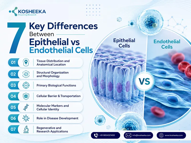
3. Primary Biological Functions
Epithelial cells are responsible for upholding barrier mechanisms that aid in sensory perception, absorption, secretion and filtration of waste products. For instance, in the gastrointestinal tract, epithelium is responsible for nutritional absorption from food, in the respiratory tract, they secrete mucus that traps pathogens or neutralizes toxins, while glandular epithelium is responsible for the release of proteins, hormones and water. Similarly, transitional epithelium lines the urinary tract and facilitates expansion of the bladder.
Endothelial cells actively regulate the vascular system. They form the vascular lining, control vascular tone, regulate blood coagulation, contribute to angiogenesis, and contribute to immune cell trafficking. Endothelium communicates with blood cells and its surrounding tissues. They maintain vascular integrity and respond to physiological demands. Endothelium controls vasoconstriction and vasodilation of the blood vessels, manages fluid movement, and prevents thrombosis.
4. Cellular Barrier & Transportation
Epithelium forms highly selective interfaces that separate the internal body from the external environment. Tight junctions between epithelial cells regulate ion flow, nutritional gain and microorganism elimination, while preserving tissue integrity.
The endothelium barrier is responsible for the exchange of substances between blood vessels and surrounding tissues. In the healthy state, the endothelium maintains permeability that enables adequate flow of fluids, oxygen, metabolites, or signalling molecules. Simultaneously, they maintain permeability enough that it restricts toxins or other substances from entering the blood. The transport mechanism involves passive diffusion, active transportation, receptor-mediated fluid uptake, or transcytosis.
Both endothelium and epithelium facilitate selective transportation that aids in maintaining adequate organ functions. Selective transport is essential for maintaining organ function.
5. Molecular Markers and Cellular Identity
The molecular markers distinction enables the identification of specific cells. Epithelial cells express cytokeratins (intermediate filament proteins that contribute to cellular stability and organization of the tissue. other molecular markers includes E-cadherin (E-cad) and epithelial cell adhesion molecule (EpCAM)
The molecular markers involved in endothelial cells include CD31 (PECAM-1), VE-cadherin, von Willebrand factor (vWF), and vascular endothelial growth factor receptors (VEGFRs). These molecular markers are responsible for vascular signalling, maintenance of blood vessels, and cell adhesion.
Researchers identify these molecular markers for verification of the cell identity, purity, differentiation and authenticity. They play a crucial role in molecular fingerprinting that enables the distinguishing of cellular populations. Accurate identification of molecular markers enables authentic experimentation outcomes, reproducibility, disease modeling or development of cell-based therapies.
6. Role in Disease Development
Disruption of adequate Epithelial vs Endothelial Cells leads to specific diseased states. Epithelial cell dysfunction is related to barrier integrity, inflammation, and degeneration of tissue. In humans, 80-90% of the carcinomas originate from epithelial cells.
Endothelial dysfunction acts as a major hallmark of cardiovascular disease and inflammatory conditions. The impairment of endothelial signalling leads to abnormal blood clotting, high blood pressure, vascular inflammation and metabolic disorders like diabetes or chronic inflammatory bowel disease.
Researchers explore cellular dysfunction for early stage disease identification. The detailed understanding of healthy cellular functioning and dysfunction enables development of valuable insights for disease mechanisms and therapeutic development.
7. Regenerative and Research Applications
Researchers widely explore both epithelial and endothelial cells in regenerative research. Epithelial cells, including kidney epithelial cells, are widely used for tissue regeneration, development of an organoid system, evaluation of drug toxicity or investigation of organ-specific diseases.
Endothelial Cells are implicated in vascular engineering, the development of angiogenesis, the development of vascular grafts, and the improvement in implanted tissues.
Epithelial vs Endothelial Cells: At a Glance
Table: Difference between Endothelium and Epithelium
| Features | Epithelial Cells | Endothelial Cells |
| Location | Cover body (skin), lines major organs or cavities, exposed to outside environment | Lines interior of blood, lymphatic vessels and heart |
| Function | Sturdy barrier (protection, adsorption or secretion) | Tissue homeostasis, blood flow, vascular permeability, immune response |
| Structure | Tightly packed can be single layered or stacked | Single thin layer, flat cells, |
| Cytoskeleton | supported by keratin filaments | supported by vimentin filaments |
| Markers | Cytokeratins, E-cadherins, EpCAM | CD31, VE-cadherin, vWF, CD34 |
| Research Application | EMT studies | Vascular biology, angiogenesis, drug delivery, blood-brain barrier |
| Diseases | Carcinoma, cystic fibrosis, asthma | Atherosclerosis, hypertension, diabetic retinopathy |
Conclusion
Endothelium vs epithelium understanding is crucial in identifying distinct cell types. The identification of the cellular identity for researchers enables them to use adequate cells for research. Various biomedical research, including drug toxicity, disease modeling, screening of novel compounds, molecular pathways, or regenerative medicine applications.
FAQ’s
Q- What is the Main Difference Between Endothelium and Epithelium?
Endothelium makes the lining of the blood and lymphatic vessels, as well as the heart. Epithelium forms the outermost layer of the body and major organs.
Q- Where are Endothelial Cells Found in the Body?
Endothelium is found in arteries, veins, capillaries and lymphatic vessels.
Q- How Do Researchers Distinguish Epithelial vs Endothelial Cells in Culture?
Differences between endothelium and epithelium are determined based on molecular marker availability and cytoskeletal structure.
