Introduction
The liver is the critical organ in the body responsible for an array of functions. It regulates metabolism, nutrient storage, detoxification, and protein synthesis. In biomedical research, hepatic function investigation plays a crucial role in understanding liver biology. Mouse Hepatocytes are an essential tool for investigating various liver diseases, drug metabolism, hepatotoxicity, metabolic disorders, fibrosis, cirrhosis, and liver cancer. The current advancement in developing 3D culture models enables the investigation of regenerative medicine and liver disease with greater accuracy. Mouse hepatocyte-based platforms enable bridging fundamental liver biology with advanced translation and regenerative research.
Mouse Hepatocytes: Foundation for Liver Research
Biological Function
Hepatocytes are predominant parenchymal cells of the liver that account for 70-80% of its cellular mass. The cells are highly specialized, consisting of hepatic lobules, sinusoidal endothelial cells, Kupffer cells, and hepatic stellate cells. Together, these cells maintain liver homeostasis and metabolic functions like protein and carbohydrate metabolism. These are crucial sites for xenobiotic metabolism mediated via the activity of P450 enzymes and other detoxification pathways.
Biomedical Research Relevance
Mouse hepatocytes shares a closer biological relevance with humans. Researchers use mouse hepatocyte cells for understanding normal physiology, diseased conditions, identification of diagnostic biomarkers, and development of targeted therapeutics. The major advantages include:
- Genetic tractability of the hepatocytes in the mouse liver allows researchers to manipulate specific genes and investigate their role in hepatic development, metabolism, or disease progression
- Consists of various conserved pathways like lipid metabolism, oxidative stress response, drug biotransformation, and liver regeneration
- Facilitates undertaking translational liver research, supports non-alcoholic fatty liver studies, drug-induced liver injury, fibrosis, cirrhosis, hepatocellular carcinoma, etc. [1]
Mouse Hepatocyte Culture Platforms
Researchers use mouse hepatocytes culture platforms as per the experimental designs and needs. Various platforms include:
Primary Mouse Hepatocyte Culture
- It involves the isolation and characterization of hepatocytes from distinct mouse models
- Common mouse model includes FVB/N, C3H/He, C57BL/6
- Characterization via determination of biological markers: e.g., Fas receptors, Bak/Bax/Bid protein, Caspase-3/7/8
- Morphologically, the cells are polygonal with distinct granular cytoplasm, a prominent border, and binucleated
- Genetic Markers: Albumin, Glucose-6-phosphatase, Serpina 1
- Protein Markers: Albumin Secretion, Asialoglycoprotein receptor
- Metabolic Markers: Drug metabolism (Cytochrome P450), Urea synthesis [1]
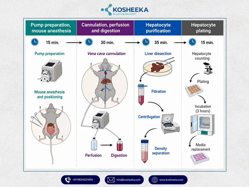
Isolation of Mouse Hepatocytes
Mouse Liver Cell Lines
- Secondary cell culture or mouse liver cell lines are crucial in vitro research models
- Common Mouse Liver Cell Lines includes: Non-tumourigenic (AML-12, FL83B), and Tumorigenic (Hepa1-6, BNL CL.2)
- Suitable research model for large-scale studies, and development of preclinical data
- An efficient model for gaining high reproducibility and high-throughput studies
Advanced Three-Dimensional Culture Systems
- Development of 3-D model (Spheroids/ Organoids)
- Suitable model bridge between 2-D flat culture (cell line culture) and in vivo model (animal studies)
- Mimics complex tissue microenvironment efficiently
- Reduces the need for animals in research
Applications in Drug Discovery and Toxicology
Hepatic Drug Metabolism Studies
- Investigation of the metabolic fate of the therapeutic compounds
- Primary site for Xenobiotic metabolism
- Express a wide range of drug metabolism (CYP50 family)
- Prediction of drug-drug metabolism, metabolic profiling of the therapeutic compound
- Determination of a drug’s ADME properties (absorption, distribution, metabolism, elimination)
- Determination of the pharmacokinetic properties of a new drug prior to clinical testing
Hepatotoxicity Assessment
- Hepatocytes in the mouse liver enable the development of a controlled platform for the evaluation of cellular response against therapeutic compounds.
- Enable evaluation of cell viability, mitochondrial dysfunction, oxidative stress, inflammatory signalling, apoptosis pathways, or alteration of liver-specific biomarkers.
- Evaluation of drug-induced liver injury (DILI). This is crucial in drug withdrawal or clinical trial failure.
- Determination of hepatotoxicity liabilities enables establishing safety assessment and reducing the risk of late-stage drug attrition [2]
Predictive Models for Pharmaceutical Research
- Crucial predictive model for the evaluation of drug efficacy and the safety of novel therapeutic compounds
- High-throughput evaluation of large compound libraries
- Screening of therapeutic compounds with desirable metabolic and pharmacological properties
- Cost-effective and experimentally controlled approach
- Development of 3-D experimental model- enable 3Rs (Replacement, Reduction, and Refinement)
- Significant reduction in animal testing dependence
*Drug-induced liver injury is among the major causes of drug attrition. This makes mouse hepatocytes the leading components of preclinical research
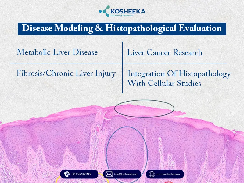
Disease Modeling & Histopathological Evaluation
Metabolic Liver Disease
- Modeling distinct liver diseases, e.g, Metabolically-dysfunction-associated steatohepatitis (MASH), Non-alcoholic fatty liver disease (NAFLD)
- It represents a pathological condition of Liver disease: elevated fatty acids, glucose, lipotoxic compounds, oxidative stress, mitochondrial dysfunction, and impaired insulin signalling
Fibrosis/Chronic Liver Injury
- Identification of inflammation, excessive extracellular matrix deposition
- Examination of early cellular events leading to fibrogenesis (hepatocytic stress, apoptosis, release of pro-inflammatory mediators)
- Interactions between hepatocytes and other liver-resident cells (hepatic stellate cells, macrophages)
Liver Cancer Research
- Mouse hepatocyte-based models are valuable tools for hepatocellular carcinoma (HCC)
- Investigation of genetic, epigenetic, and metabolic alterations involved in tumour initiation, proliferation, or progression
- Diagnostic biomarker identification for cancer progression
- Development of a targeted treatment approach
Integration of Histopathology with Cellular Studies
- Mouse Liver Histopathology allows identification of structural alterations
- Inflammation, steatosis, necrosis, and fibrosis identification
- Correlation of in-vitro observation and histopathological conditions enables identification of cellular and molecular changes [2]
*Kosheeka, India is a renowned laboratory that supplies primary and secondary cells for various research purposes including liver diseases.
Mouse Embryonic Stem Cells: Regenerative Research
- Regenerative research is rapidly emerging for multiple chronic conditions, including liver disease
- Mouse embryonic stem cells are pluripotent in nature, which allows for studying liver developmental biology
- Establishing directed differentiation strategies
- Understanding the key signalling pathway regulating hepatic lineage commitment
- Advancements in establishing tissue engineering and liver repair research
What are the Current Challenges in Mouse Hepatocyte Models in Research?
- Maintaining functional stability in primary mouse hepatocytes is a complex and tedious task
- Replicating the native liver microenvironment while using a mouse liver cell line is not guaranteed
- Discrepancies in translating preclinical results into human studies due to interspecies differences
- Discrepancies in genetic makeup, physiology, anatomical structure, and immunological response limit the experimental translation
- 3-D model (Organoids or Liver-on-a-Chip Systems) lacks complex human systems like vascular or biliary network, batch-to-batch variability, cellular heterogeneity, and high cost
Did You Know?
- Certain human-specific viruses (Hepatitis B/C) cannot infect mice, so researchers have developed a ‘humanized chimeric mouse’ model
- Researchers maintain a self-renewing Zone 2 mouse hepatocyte cell population to support continued regeneration
- On isolation of mouse hepatocytes, the protein profile rapidly changes (does not undergo apoptosis rapidly). A scientist exploits the opportunity to unveil how tissue damage evolves in the body.
Conclusion
Mouse hepatocyte cultures have a wide range of applications in biomedical and regenerative research. Using a mouse model enables researchers to develop preclinical data related to diagnosis and therapeutics. Their smaller size, rapid reproduction, and physiological similarities (99% genomic similarity with humans) make them suitable research models. Researchers must focus on maintaining uniform strains and avoiding overbreeding to maintain the high credibility of the research outcome.
References
- Charni-Natan M, Goldstein I. Protocol for primary mouse hepatocyte isolation. STAR protocols. 2020 Sep 18;1(2).
- He L, Pu W, Liu X, Zhang Z, Han M, Li YI, Huang X, Han X, Li Y, Liu K, Shi M. Proliferation tracing reveals regional hepatocyte generation in liver homeostasis and repair. Science. 2021 Feb 26;371(6532):eabc4346.
FAQ’s
Q- What are Mouse Hepatocytes?
Mouse hepatocytes are the primary liver cells that make upto 80% of the liver volume. These cells play a crucial role in liver research and regenerative medicine.
Q- How Do Mouse Liver Cell Lines Differ from Primary Cells?
Primary hepatocyte cells are isolated directly from the mouse liver tissue. They share physiological properties with the organism (genetic makeup, physiological condition, chromosomal number). Mouse live cell lines are transformed cells that have a longer lifespan. They are suitable for large-scale studies and for gaining data reproducibility.
Q- Why is Mouse Liver Histopathology Important?
This enables researchers to gather evidence related to cellular-level evidence of liver damage, treatment efficacy, and disease progression.
Q- Can Mouse Embryonic Stem Cells Become Hepatocytes?
Yes, mouse embryonic stem cells are pluripotent in nature and can differentiate into completely functional hepatocytes or liver cells.


The focus on liver-derived primary cells highlights how important physiologically relevant models are for studying metabolism, toxicity, and disease mechanisms. It would also be interesting to see more discussion around how researchers balance the advantages of primary hepatocytes with challenges such as donor variability and maintaining cell function over time.