Dendritic cells are immune cells with unique features of their own. They possess the ability of cross-presentation. They can bridge the innate and adaptive arms of the immune system. Their crucial role in the immune response has implicated them in autoimmune diseases and cancer. Scientists are conducting high-throughput research into understanding the underlying signaling pathways for such pathologies and developing therapeutics involving them. In this blog we will discuss their immunobiology.
Dendritic Cells
In 1973, two Canadian scientists, Ralph Steinman and Zanvil Cohn, discovered dendritic cells (DCs) in mouse spleen and peripheral lymph nodes. They named them dendritic based on their cytoplasmic extensions. DCs originate from the hematopoietic stem cells in bone marrow. They are present throughout the body- blood, lymphoid tissues, and non-lymphoid tissues.
Functions of Dendritic Cells
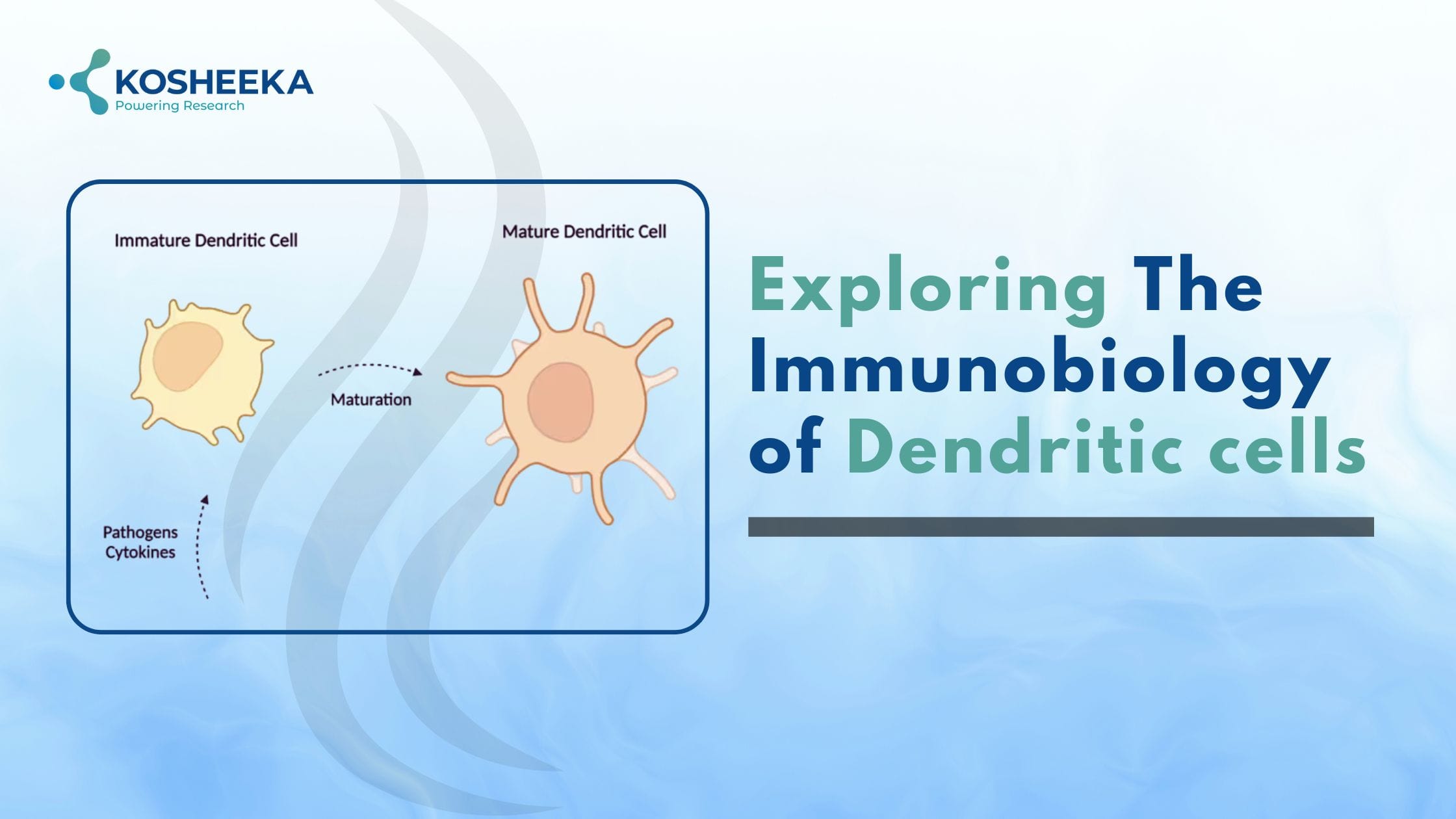
DCs are the most potent antigen-presenting cells (APCs) to T lymphocytes. They perform several vital tasks in the immune system response (Fig 1).
Immune Response
DCs serve as APCs to T lymphocytes and also interact with B lymphocytes, and macrophages. The hematopoiesis process in bone marrow produces immature DCs. They migrate to the non-lymphoid tissues to monitor for pathogenic antigens. Their surfaces contain pattern recognition receptors (PRRs). The PRRs recognize the pattern-associated molecular patterns (PAMPs) on the pathogen surface, which are evolutionarily conserved regions of pathogens. After recognizing the antigen, DCs uptake the antigen and undergo maturation. They migrate to the lymph nodes and lose their antigen uptake ability. To transform into APCs, they express major histocompatibility complexes (MHC) I and II along with costimulatory molecules CD40, CD80, and CD86. Mature DCs present the antigen to T lymphocytes, stimulating the production of IL2 cytokine. It, in turn, causes the T lymphocyte expansion, B lymphocyte development, and antibody production.

Immune Tolerance
DCs also maintain immune homeostasis by presenting self-antigens to T lymphocytes, thus also contributing to immune tolerance. There are two kinds of immune tolerance- central and peripheral. In central tolerance, DCs participate in the elimination of T lymphocytes that bind to self-antigens with high affinity. This process of selection occurs in the thymus. Furthermore, in the absence of infection, immature DCs monitor the environment for self-antigens such as cell debris, apoptotic cells, etc., as well as cells that are present in later stages of life. These cells do not have access to the thymus. Therefore, DCs capture and process these antigens, contributing to the peripheral tolerance.
Cross-Presentation
DCs have the unique feature for cross-presentation. Generally, APCs present an endogenous antigen on MHC I to CD8+ T lymphocytes. In contrast, CD4+ T lymphocytes recognize an exogenous antigen on MHC II. However, a DC has the capacity for cross-presentation of exogenous antigens on MHC I molecules to CD8+ T lymphocytes.
Heterogeneity of Dendritic Cells
One of the puzzling aspects of DCs is their heterogeneity. Scientists have observed variation in these cells depending on their origin and anatomical location. For example, they show considerable differences when derived from lymphoid and myeloid precursors. Myeloid DCs typically reside in the marginal zone of the spleen. On the other hand, lymphoid DCs are present in regions of the spleen with a high concentration of T lymphocytes. They also vary in their surface expression of CD11c markers.
Types of Dendritic Cells
The DC heterogeneity has led to their categorization into different subsets. These subsets are present in circulation and in tissues.
Conventional or Classical dendritic cells (cDCs)
They further have two subpopulations. In mice, these populations are identified by their expression of CD8α and CD103, or CD11b. The population with CD8α or CD103 expression demonstrates the unique cross-presentation ability. Additionally, CD8α positive cDCs can also express CD1d glycolipid and polarize natural killer T lymphocytes to form helper T lymphocytes or Th2 cytokines. cDCs with CD11b expression are the most abundant DCs in the lymphoid tissues. The production of IL6 and IL23 is also a defining characteristic of CD11b positive cDCs.
Plasmacytoid dendritic cells (pDCs)
They originate from lymphoid precursors. Due to their ability to differentiate into cDC, their amount varies in the residing tissue. They have a round shape and distinct plasmacytoid morphology similar to the lymphocytes. They also contain a secretory compartment. These dormant cells produce high amounts of interferon (IFN) γ upon stimulation by toll-like receptors (TLR) 7 and TLR9. They play a crucial role in pathogen infections, and in the absence of infections, they display immune tolerance. Owing to the expression of MHC II and costimulatory molecules, pDCs can serve as APCs.
Monocyte-derived dendritic cells (moDCs)
The moDCs differentiate from myeloid precursors. The monocytes circulate in blood and express receptors for growth factors, cytokines, and chemokines. These receptors drive the monocyte migration towards the site of infection or inflammation. Subsequently, they differentiate into moDCs and release tumor necrosis factor α (TNF α) and inducible nitric oxide synthase (iNOS). These molecules grant moDCs innate protection and therefore also refer to them as TNFα, iNOS-producing DCs. They are highly potent in antigen processing, presentation, and cross-presentation. Although moDCs and cDCs with CD11 positive subset share similar markers, moDCs can be distinguished by the expression of Fc-gamma receptor I (FcgRI) and CD64.
Clinical Relevance
Transplantation: Graft vs. host disease (GVHD) is a typical complication of transplantation. Here the graft or the administered cells mount an immune attack on the recipient or the host. Host DCs initiate this process by presenting the host antigen to the T lymphocytes present in the graft. In the case of organ transplantation, the graft DCs present the graft antigen to host T lymphocytes. GVHD eventually results in transplant failure and a high mortality rate.
Autoimmune Diseases: DCs are an integral part of immune tolerance. Several genetic and environmental factors can create an imbalance in their tolerogenic response. For example, in systemic lupus erythematosus, pDCs release high amounts of IFNα that activate the cDCs, disrupting their ability for peripheral tolerance. DCs are also associated with the pathogenesis of early diabetes type I. Due to their role in maintaining regulatory T lymphocytes, it has been suggested that type I diabetes can be treated by increasing DC number.
Cancer: T lymphocytes play a huge role in immunity against tumor cells. The cDCs activate these cells by endocytosis of tumor-associated antigen, migrating to lymph nodes, and activating T lymphocytes. Their ability to present both exogenous and endogenous antigens renders them suitable for promoting T cell response and polarization. However, tumor cells produce cytokines such as IL6, IL10, and vascular endothelial growth factor (VEGF) that suppress the DC differentiation and activation.
Dendritic Cell-Based Therapy
DCs can potentially activate adaptive immune cells that can in turn activate the innate immune system. Thus, they link both the innate and adaptive immune system. These characteristics have prompted investigations into DCs for therapeutic purposes. They can enhance the immunogenicity of the vaccines by activating the adaptive and innate immune system. DC-based vaccines have been developed and proved to be safe and effective. The DC progenitors are cultivated in the lab and differentiated into DCs by cytokine addition. These cells are loaded with tumor antigens via different techniques like electroporation or vector-based methods. After infusing these cells into the patient, they migrate to lymph nodes and present the antigen to naïve T lymphocytes, thus stimulating an anti-tumor immune response.
Product-Related Queries, Or Partnership Inquiries
Conclusion
DCs are essential for establishing immune response and tolerance. Therefore, they have implications in autoimmune disorders and other pathological conditions. Research into delineating different subsets of DCs, their functions, and therapeutic applications is ongoing. Kosheeka delivers DCs from diverse species to assist in your research endeavors. Our team provides high-quality dendritic cells with assured viability, purity, and functionality.
FAQs
Q: What are the functions of dendritic cells?
Dendritic cells act as antigen-presenting cells to T lymphocytes. They also eliminate T cells that interact with self-antigens, thus contributing to immune tolerance.
Q: What are the unique features of dendritic cells?
Dendritic cells are unique in their ability for cross-presentation. They can present an exogenous antigen to CD8+ T cells (they recognize endogenous antigens). They also link adaptive and innate immunity.
Q: How do dendritic cells cause graft vs host disease?
Host dendritic cells present host antigen to transplanted graft T cells, causing the graft T cells to mount an immune response against the host. The process results in graft vs host disease.
Q: What are the different disorders dendritic cells are involved in?
Dendritic cells are implicated in autoimmune disorder and cancer due to their significant role in immune tolerance and T cell activation.

Really great breakdown of what dendritic cells are and how vital they are for our immune system. The way they act as a bridge between innate and adaptive immunity, their ability to present antigens, and especially their role in “cross-presentation” makes them key players. Also interesting to learn how their subtypes (conventional, plasmacytoid, monocyte-derived) behave differently depending on where they are, and how that contributes to both immune response and immune tolerance.
One point that stands out is their therapeutic potential—whether for vaccines, treating autoimmunity, or cancer—because manipulating dendritic cell function could shift the immune system in beneficial ways. Thanks for shedding light on this complex but exciting area!
Thank you so much for your thoughtful comment. I completely agree with your point on the therapeutic potential of dendritic cells. Research into dendritic cell–based immunotherapies is one of
the most promising areas right now, especially in cancer and atherosclerosis. It will be fascinating to see how future discovery and research will shape their clinical applications.