
Keywords: Primary Culture, Skin Culture, Keratinocytes
The skin protects us from the external environment and is composed of 2 layers: the inner dermis and the outer epidermis. The epidermis forms what is called an “an intact epidermal barrier”; this separates our body from the various unwanted molecules outside such as UV rays, pathogens or allergens.
The major cell type of the epidermis is the keratinocyte; 95% or more of the cell mass is composed of keratinocytes. The major function of the keratinocytes is to form an intact barrier to the external surroundings by maintaining the structural integrity of the epidermis. Another function of keratinocytes is to function in the innate immune response against pathogens as the first line of defence in the skin. Keratinocytes can activate the immune system by cytokines including IFNβ, IL6 and TNFα that are involved in inflammation, chemokines, as well as, antimicrobial peptides when exposed to external stimuli. These stimuli include pathogens or molecules called damaged-associated molecular patterns (DAMPs) that are produced when injured or exposed to UV. These secreted molecules can recruit or activate immune cells. It is very important that there is a balance in this immune activation-if unchecked, the result can be auto-inflammatory skin diseases.
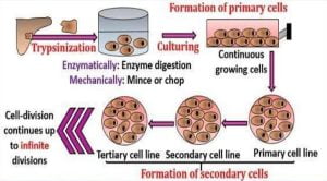
An article published in the Journal of visualized experiments in 2017 by Li and team reported the use of a Primary Cell Culture of keratinocytes to study skin biology. The team used tail skin to isolate keratinocytes. When the dorsal skin of mice is at the telogen phase or resting phase of the hair cycle, there are 1-2 layers of cells in the epidermis that makes the separation of this layer from the dermis difficult resulting in very low cell yields. The difficulty increases due to the presence of excessive hair on the dorsal surface.
The washed skin is digested sequentially with dispase and trypsin followed by washing and seeding in medium containing defined growth supplements. The cells showed differentiation when Calcium levels were increased as seen in vivo. There was a change in morphology of the cells and remodelling of the protein actin to cause what is called stratification. When the keratinocytes were exposed to UVB, a cytokine called TNFα was released that is involved in inflammation showing the role of keratinocytes in immune activation.
Thus, primary keratinocyte cultures serve as valuable in vitro tools to represent these cells that form the barrier and also activate the immune system. This can provide valuable insights into the field of the many “layered” skin biology.
References:
Johansen C. (2017). Generation and Culturing of Primary Human Keratinocytes from Adult Skin. Journal of visualized experiments : JoVE, (130), 56863. doi:10.3791/56863.
Li, F., Adase, C. A., & Zhang, L. J. (2017).Isolation and Culture of Primary Mouse Keratinocytes from Neonatal and Adult Mouse Skin. Journal of visualized experiments :JoVE, (125), 56027. doi:10.3791/56027.

