Apart from the usual functions of the kidney listed in high school textbooks, it is targeted by many xenobiotics and also develops tumors. According to research published in Clinical genitourinary cancer in 2019 by Saad and team, 65,340 cases of the most common renal cancer called renal cell carcinoma (RCC) was diagnosed in the US in 2018 with the estimated mortality figure pitched at 14,970. It has been described as the 6th cause of malignancy in males and the 10th in the case of females. RCC is of several types on the basis of histology- 80 to 85% of renal cell tumors are conventional or clear cells originating from the proximal tubular epithelium. 10% is papillary RCC while 5% is accounted for by chromophobe RCC primary cultures.
In order to assess the effects of toxicants or the mechanisms of development of tumors to thereby develop a therapeutic system require appropriate in vitro models. This becomes especially relevant given that the precise causes of RCC are yet to be known in detail. The disease also shows resistance to the traditional therapeutic approaches such as chemo or radiotherapy. Additionally, testing new chemical molecules for their ability to target cancer also requires healthy cells. This can highlight possible toxic effects.
Among the various cells, a significant portion of the cortex is human proximal tubular epithelial cells (HPTEC). These cells are targeted by metals, antibiotics, and other drugs. The challenge with using immortalized Cancer Cell Line is their altered characters that may not truly recapitulate the actual in vivo tissue.
In order to study the processes associated with drug-induced damage in vitro, primary cultures make their entry. These systems while closely being representative of the in vivo tissue also are not challenged by the interferences seen while assessing tissues. In order to specifically isolate HPTECs, approaches such as Percoll centrifugation or microdissection have been reported. However, these face challenges in terms of low yield and the amount of time and labor involved.
A team led by Valente published a 60-minute protocol in PlosOne to culture HPTEC from kidney tissue samples. Once identified, the samples were processed to remove the capsule and adjacent medulla followed by ice-cold HBSS washes. Chopped tissue was mixed with collagenase in medium followed by sieving through 100 µm, 70 and 40-µm filters. The media recommended are DMEM/F-12 and GlutaMAX-I™ supplemented with 10% heat-inactivated fetal bovine serum (FBS), penicillin/streptomycin, fungizone, and human transferrin. Human renal tumor cells (HRTC) can also be cultured on RPMI 1640 medium with supplements.
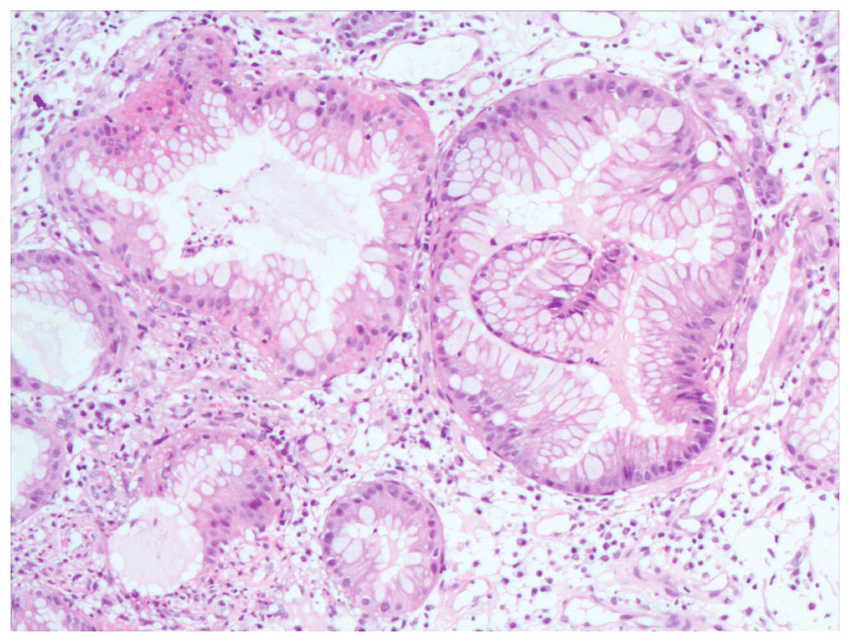
There were no fibroblasts detected in the primary cultures and the cells showed the expected morphology of cobblestone appearance and domes. The cells also expressed the epithelial cell marker cytokeratin and the proximal tubular marker called aminopeptidase A. RCC primary cultures too displayed a mixed morphology that is in agreement with the heterogeneous nature shown by research.
Thus, such primary cultures of the kidney cells can do away with the use of animal models that not only have ethical issues but also variations across species. Apart from studying tumors and testing potential drugs, the use of permeable membrane filter supports can allow a closer representation of actual in vivo tissue and the transport of drugs across the proximal tubule. This can hence propel science towards realizing the goals of addressing serious diseases such as cancer that threaten our very existence.
References:
Saad, A. M., Gad, M. M., Al-Husseini, M. J., Ruhban, I. A., Sonbol, M. B., & Ho, T. H. (2019). Trends in Renal-Cell Carcinoma Incidence and Mortality in the United States in the Last 2 Decades: A SEER-Based Study. Clinical genitourinary cancer, 17(1), 46–57.e5. DOI:10.1016/j.clgc.2018.10.002
Valente, M. J., Henrique, R., Costa, V. L., Jerónimo, C., Carvalho, F., Bastos, M. L., … Carvalho, M. (2011). A rapid and simple procedure for the establishment of human normal and cancer renal primary cell cultures from surgical specimens. PloS one, 6(5), e19337. DOI:10.1371/journal.pone.0019337


