
The growth of normal human mammary epithelial cells, including luminal, myoepithelial, and/or basal cells, is tightly controlled. Mammary epithelial cells grow for a finite span and eventually die or undergo senesce. Human, rat, and murine mammary epithelial cell have provided evidence that essential initial steps in mammary carcinoma cell line growth involve the loss of senescence checkpoints for encouraging immortalization. In addition, mammary epithelial cell culture model systems have identified a number of genes whose alterations are involved in mammary carcinoma cell line development. Additional insights come from using transgenic overexpression of carcinoma-promoting genes or deletion of cancer suppressor genes. Let us get some insights into mammary epithelial cells and their cancer progression.
Mammary Epithelial Cells
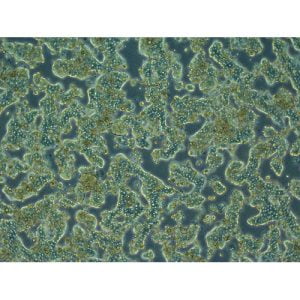
The mammary gland consists of a branching ductal system that ends in terminal ducts with their associated acinar structures (terminal ductal-lobular units or TDLUs), along with interlobular fat and fibrous tissue. Histological examination of the TDLU has shown two major types of cells: inner secretory luminal cells and outer contractile myoepithelial cells. Two types of luminal cells are present lining the mammary gland ducts and alveoli. In addition to these, there is also evidence regarding the presence of stem cells and progenitor cells for mammary epithelial cells.
For more than two decades, researchers have attempted to develop mammary epithelial cell culture models that resemble human breast cancers in vivo. In order to establish such models, culturing non-cancerous mammary epithelial cells was necessary using a human mammary epithelial cell growth medium. To design an optimum growth medium, researchers prepared a defined medium DFCI-1 to culture mammary epithelial cell and epithelial carcinoma cell lines but there was difficulty in establishing primary carcinoma cell culture.

Cultures derived from reduction mammoplasty or mastectomy specimens exhibit considerable heterogeneity but researchers devised ways to establish mammary epithelial cell from these specimens. In the procedure, the tissue is finely chopped, digested by collagenase and hyaluronidase, and plated as organoids. Over a week, multiple types of epithelial cells and fibroblasts are seen but fibroblasts are removed by differential trypsinization, leaving mammary epithelial cells. For information on mammary epithelial cell growth medium or serum-free cell culture media, click here
Mammary Epithelial Cell Carcinoma
Mammary carcinoma exhibit both inter-and intra-tumoral heterogeneous nature. Several study reports have indicated that human mammary epithelial cell cancers exhibit diverse phenotypes according to pathological features and therapy response. Studies have identified distinct gene profiles to classify mammary carcinoma cell lines. Five categories of mammary carcinoma cells can be described as a basal epithelial-like group, ErbB2-overexpressing group, normal mammary epithelial cell-like group, luminal epithelial cell type A, and luminal epithelial cell type B. Importantly, these molecular classifications provide a strong rationale for studying various mammary epithelial cell subtypes and models to understand carcinoma cell line molecular diversity.
Further gene expression profiling shows that within each subtype, tumors can exhibit further variability in gene expression and drug susceptibility, making sense of distinct patient complications. Molecular profiling studies report that the gene expression patterns of cancer subtypes align with normal mammary epithelial cell lineages and this suggests that tumor subtypes may originate from distinct mammary epithelial cell subpopulations. It is widely believed that mammary epithelial stem cells/progenitor cell populations may serve as carcinoma initiating cells since their longevity and self-renewal ability can afford genetic mutation accumulation. For more information on mammary epithelial cell carcinoma and its hierarchy, click here
If your lab is working on mammary epithelial cell carcinoma, Kosheeka can help you procure the best quality of human primary cell culture, tissue-specific primary cells, and disease-specific primary cells. Contact info@kosheeka.com for further inquiries.


Great Insight On Mammary Epithelial Cells And Carcinoma, looking for more updates.
#Kosheeka #CellCulture
Pingback: Kosheeka (kosheeka) | Pearltrees