Introduction
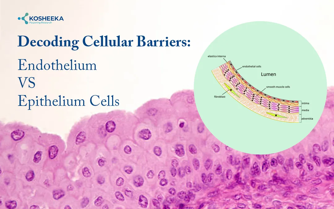
Beneath every multicellular organism lives a cellular divide- endothelial vs epithelial cells subtly design pathology and physiology.
Endothelium vs Epithelium Cells are two distinct tissue/cell types that cover your body surface. The location and function are distinct. Endothelium cells coat the inner lining of the blood and lymphatic vessels. This cellular layer maintains tissue homeostasis. In contrast, epithelium cells line the major organs and cavities in the body. These cells/tissues interact with external environments like skin, digestive system and lung. They act as major barriers that defend the body against environmental factors.
In molecular biology research, both epithelial vs endothelial cells play crucial roles in understanding cellular mechanisms, molecular pathways, disease diagnosis, therapeutics, translation research, and regenerative medicine.
Difference Between Endothelium vs Epithelium
The functional difference between endothelium vs epithelium are distinct. This includes:
| Features | Endothelium | Epithelium |
| Location | Lines inner lining of the blood, lymphatic vessels, and heart | Forms outer lining of the body external and internal major organs |
| Morphology | Flattened, elongated structure | Distinguished shaped depending on its location and function |
| Cellular Layers | Single layered | Multiple layers depending on location |
| Function | Tissue homeostasis, regulates vascular tone, blood clotting, immune regulation Facilitates gaseous exchange | Forms protective barrier, facilitates absorption, sensation, and secretion Protects organism from any external injury or environmental stressors like dehydration and pathogens |
| Secretory Function | Secretes various molecules like prostacyclin, nitric oxide, endothelin, etc. | Absence of Secretary function |
| Disease Association | Cardiovascular disease | Skin, lung, kidney disease, etc. |
| Basement Membrane | Unavailable | Anchored with a basement membrane for structural supports |
| Origin | Mesoderm derived | Ectoderm, endoderm, or mesoderm derived, differentiate at the time of embryonic development |
| Regenerative Capability | Limited regenerative capability | Rapid regenerative ability, maintains tissue integrity |
Functional Divergence in Endothelium vs Epithelium Cells
Functionally, both epithelial and endothelial cells have distinct functional features. This includes:
- Endothelial Cells: Responsible for the regulation of vascular tone. Mediates via nitric oxide signaling and maintains tissue homeostasis. Endothelial cells modulate cellular permeability at the time of inflammation, tissue repair mechanisms, angiogenesis, vasculogenesis, metabolic and endocrine function. These cells are major barriers or protectors against pathogens, oxidative stress, and regulate vascular integrity at the time of pathological or physiological disturbance [1,2].
- Epithelial Cells: These cells have an active role in the barrier mechanism and transportation. Epithelial Cells have selective physical barriers with tight junctions. It regulates paracellular transportation and forms a semipermeable interface between blood and tissues. They have an active role in fluid exchange, exchange of solutes, and cell function. Another major function is to facilitate filtration, secretion and absorption. For instance, renal epithelium mediates filtration and reabsorption, intestinal epithelium supports nutrient absorption, and glandular epithelium facilitates the secretion of enzymes, hormones, and mucus. The function is tissue or organ-specific [1,2].
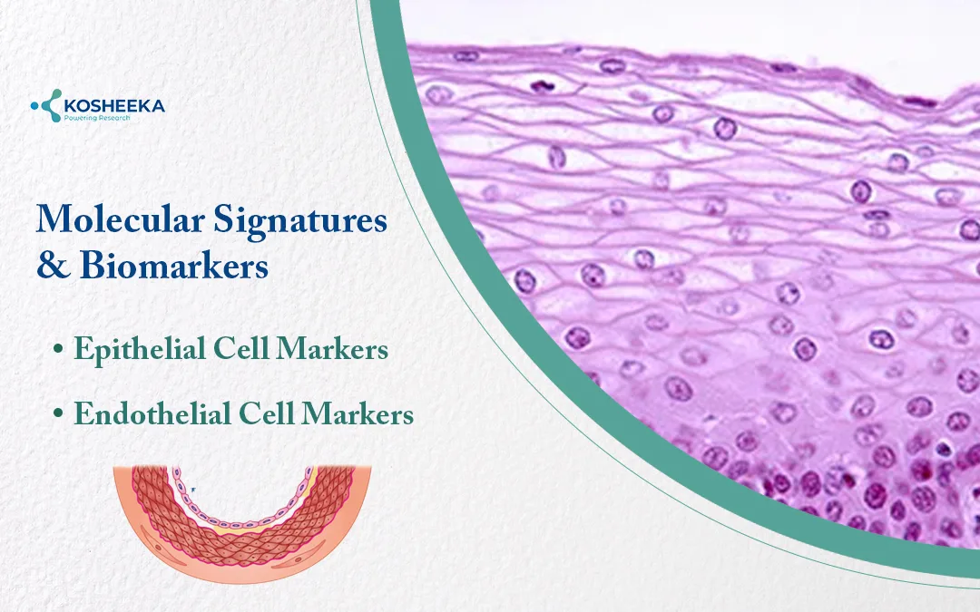
Molecular Signatures and Biomarkers
Epithelial Cell Markers
- Cytokeratins (CK8, CK18, CK19)
- E-cadherin (CDH1) facilitates cell–cell adhesion
- EpCAM acts as a surface marker
- Claudins, occludin, and ZO-1 act as markers for tight junction proteins
- Presence of tissue-specific markers (MUC1 in glandular epithelium)
- Epithelial dysfunction associated with E-cadherin and tight junction proteins alteration [2]
Endothelial Cell Markers
- CD31 (PECAM-1)
- VE-cadherin (CD144)
- von Willebrand factor (vWF)
- Acts as a receptor for various molecular signalling (VEGF-R, Tie2, TGF-β, Wnt) [1]
*These molecular biomarkers play pivotal roles in disease diagnostic, cancer biology, vascular disease and regenerative medicine. They access various molecular processes like cell identity, integrity, pathological transition, etc.
Application of Endothelium vs Epithelium Cells
The key application of epithelial cells involves protection, and maintaining secretory barriers on external/internal body surfaces. Endothelial cells are associated with vascular transportation and maintaining tissue homeostasis.
Endothelium: Endothelial Cells in Vascular Biology
- Angiogenesis and Vasculogenesis: Crucial role in new vessel formation. Vasculogenesis contributes to de novo blood vessel formation from progenitor cells. Regulates angiogenesis pathways such as VEGF, hypoxia-inducible factors, or FGF
- Endothelial Dysfunction in Disease: Associated with reduced nitric oxide bioavailability, an increase in oxidative stress, and compromised vasodilation
- Inflammation and Immune Interactions: Activates immune response, expresses adhesion molecules (ICAM-1, VCAM-1, selectins). Facilitate leukocyte adhesion and promote transmigration. Modulates inflammation, immune surveillance, or tissue injury response
Epithelium: Kidney Epithelial Cells
- Role of Nephron Structure: Kidney epithelial cells are responsible for forming distinct nephron segments (podocytes in the glomerulus). Promotes selective filtration and maintains the glomerular filtration barrier
- Maintaining Transportation Mechanisms and Ion Balance: Regulates electrolyte and fluid homeostasis via active and passive transportation (Na⁺/K⁺-ATPase, ion channels, and co-transporters)
- Renal Pathophysiology: Dysfunction contributes to acute kidney disease, proteinuria, or chronic kidney disease
Experimental Models and Research Applications
Both endothelial and epithelial cells have critical research application. This involves setting up a distinct culture and experimentation in a controlled environment. The application includes:
- In-Vitro Cell Culture Models: Enable epithelial and endothelial biology study under defined conditions. Epithelial cells lines: HEK-293, Caco-2, MDCK, HeLa; Endothelial Cell line: HUVECs, HMEC-1, ECV304
- Organoids and 3D Tissue Systems: Development of advanced 3D culture systems that mimics in-vivo conditions. Epithelial organoids include kidney intestinal, whereas endothelial organoids include network studies in angiogenesis.
- Use in Drug Screening and Toxicity Studies: Evaluation of drug absorption efficacy, toxicological profiling, efficacy, shelf life determination, safety, tolerance, etc.
- Research Application in Various fields, including oncology, cardiovascular research, renal research, regenerative medicine and tissue engineering.
*Kosheeka, India, is among the renowned laboratories that grow and maintain various endothelial and epithelial primary and secondary cells. The cells are maintained with utmost care in BCL-2 laboratories and follow GMP-certified protocol.
Emerging Trends: A Promising Frontier
The use of primary and secondary culture using endothelial and epithelial cells offers a promising path towards advanced research. This includes:
- Single-cell sequencing insights
- Regenerative medicine research with stem cell therapy and exosomes
- Understanding the intercellular mechanism
- AI and bioengineering approaches
Conclusion
Epithelium and Endothelial Cells are closely placed cells in multicellular organisms. Both cell types have distinct locations and functions. In modern molecular biology research, the use of epithelial and endothelial cell lines contributes to generating preclinical data related to disease progression, diagnosis, or therapeutics.
References
- American Type Culture Collection (ATCC). Endothelial cells – primary cells overview. Manassas (VA): ATCC; [cited 2026 Mar 23]. Available from: Endothelial cells overview
- StatPearls Publishing. Renal physiology and epithelial-endothelial interactions [Internet]. Treasure Island (FL): StatPearls Publishing; 2023 [cited 2026 Mar 23]. Available from: https://www.ncbi.nlm.nih.gov/books/NBK559063/
FAQ’s
Q- What are the key molecular markers used to distinguish epithelial vs endothelial cells?
Biomarkers play a crucial role in distinguishing epithelial from endothelial cells. This includes: Epithelial cells- cytokeratins, E-cadherin, EpCAM, etc. Endothelial cells: CD31, VE-cadherin, vWF, and VEGFR-2, etc.
Q- How do endothelial cells contribute to vascular homeostasis?
Endothelial tissue maintains vascular homeostasis by regulating blood flow, vascular permeability, and immune cell trafficking. NO is the key signaling molecule. The dysfunction of endothelial cells leads to various disease conditions like atherosclerosis, hypertension, diabetes, etc.
Q- What are the applications of epithelial and endothelial cells in in-vitro models?
In-vitro research, the epithelial and endothelial cell lines aid in various research, including drug permeability, disease modeling, toxicology profiling, cellular interaction, diagnostic markers, or therapeutics. This enables understanding various complex cellular and molecular pathways such as proliferation, angiogenesis, metastasis, and tissue regeneration.

