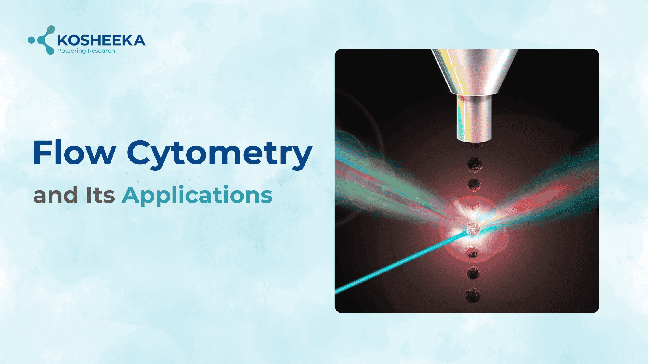
Flow cytometry is a versatile technology that simultaneously measures and analyses physical and chemical characteristics of cells/particles, as they flow in a fluid stream through a beam of light.
It provides rapid multi-parametric analysis of cells in solution (sheath fluid).
In Flow cytometer, lasers as a source of light used to produce both scattered and fluorescent light signals that are read by detectors such as photodiodes or photomultiplier tubes. These signals are converted into electronic signals and then analysed by a computer and written to a standardized format. Cell populations can be analysed and/or purified based on their fluorescent or light scattering characteristics.
Flow cytometry is a powerful tool that has applications in multiple disciplines such as immunology, virology, molecular biology, cancer biology and infectious disease monitoring. For example, it is very effective for the study of the immune system and its response to infectious diseases and cancer.
Flow cytometry allows for the simultaneous characterization of mixed populations of cells from blood and bone marrow as well as solid tissues that can be dissociated into single cells such as lymph nodes, spleen, mucosal tissues, solid tumours etc.
In addition to analysis of populations of cells, a major application of flow cytometry is sorting cells for further analysis.
How Flow Cytometry works:
A liquid suspension of cells is passed through a laser beam
![]()
The laser scatters and analyse two parameters : forward scatter (FSC) and side scatter (SSC)
![]()
Scattered light locate the cells on a cytograph
![]()
fluorescent dyes stains the cells that emit light at different wavelengths
![]()
The cell viability and other characteristics are determined by measuring degree of fluorescence
 These are the main applications of the Flow Cytometry
IMMUNOLOGY
Immunophenotyping
Flow cytometry utilizes the unique ability of flow cytometry to simultaneously analyse mixed populations of cells for multiple parameters. In immunophenotyping experiment, cells are stained with fluorochrome-conjugated antibodies that are targeted against antigens on the cell surface.
Most of these antigens are given “cluster of differentiation” numbers or CD numbers.
- Antigen Specific Responses
Cells are stimulated with specific antigen to measure Antigen specific responses and then checked for cytokine production, proliferation, activation, memory, or antigen recognition through MHC multimers.
- Intracellular Cytokine Analysis
To analyse the Intracellular cytokine cells are treated with a protein transport inhibitor (Brefeldin A or Monensin) for 2–12 hours which allows the accumulation of any cytokines within the cells enabling better detection.During this incubation cells can be stimulated with various antigens such as peptides from a vaccine to measure immune response.
- Proliferation Analysis
Several different assays and markers are used to measure cell proliferation by flow cytometry.Expression of proliferation related antigens are used as a marker for proliferation. Ki67 is expressed during cell proliferation (all phases) but not during cell quiescence.
- Apoptosis Analysis
Apoptosis, or programmed cell death, is a phenomenon that is frequently examined in immunology and other fields of study. It is used to maintain the homeostasis of the immune system by removing cells without triggering an inflammatory response.
The detection of apoptosis by flow cytometry utilizes multiple targets along the cascade of events associated with apoptosis are as below:
- Annexin V staining targets translocation of the plasma membrane.
- TUNEL Targets endonuclease digestion of DNA.
- Antibodies and dyes are used to target the activation of Caspases.
- Hoescht staining is used to assess apoptosis by determining cell cycle status and chromatin condensation in the nucleus.
MOLECULAR BIOLOGY
Fluorescent Protein Analysis
Cells are transfected with a plasmid that contains a promotor sequence and encodes for a gene of interest along with a fluorescent protein. The expression of the fluorescent protein is used as an indicator for the expression of the gene of interest.
This technology is used for multiple applications, for example in vivo tracking of transplanted cells, bacterial or viral infections, and gene knockout in cells to further elucidate gene function.
Cell Cycle Analysis
Cell cycle analysis assays consist of staining DNA with a saturating amount of DNA binding dye. The cells are fixed with a 100% methanol solution which permeabilizes the cells and then stained with the dye for analysis.
However, there are dyes that can enter living cells and stain DNA without harming the cells. In this type of analysis, samples are acquired at a low flow rate with linear amplification and then analysed using ploidy modelling software to determine the cell cycle phases.
Signal Transduction Flow Cytometry
This application uses antibodies made against resting and phosphorylated signalling molecules. The use of these reagents and specialized buffers in staining panels allows for the study of signalling pathways in mixed populations of cells.
RNA Flow Cytometry
RNA flow cytometry combines flow cytometry with fluorescent in situ hybridization (FISH) to detect RNA expression along with protein expression.
It is a useful technique when antibodies are not available for a target and RNA expression can be used instead.
Cell Sorting
Cell sorting utilizes a flow cytometer with cell sorting capabilities to separate and purify cells or particles for further analysis. Essentially, any cell or particle that can be made fluorescent can be separated by a cell sorter.
Product-Related Queries, Or Partnership Inquiries
OTHER APPLICATIONS
Absolute Cell Counting
The procedure utilizes fluorescent beads of a known concentration that is acquired along with the sample. The sample is analyzed and the gated number of cells for the population of interest is compared with the number of beads acquired in the same sample to generate the number of cells per milliliter.
Quantitative Flow Cytometry
Quantitative flow cytometry uses a bead-based standard to generate a staining curve of known fluorescence amounts. Cells are then acquired with the same instrument settings and linear regression analysis. They are used to calculate the amount of fluorescence on the cells.
Multiplexed Bead Array Assays
Multiplexed bead array assays are sets of beads coated with antibodies against specific soluble proteins or nucleic acids. Each bead has a known amount of fluorescence and a specific target which gives a location for the bead in the matrix. The collection of up to 100 beads are incubated with the sample of interest, treated with a fluorescence reporter and then acquired on a flow cytometer with at least 2 lasers to detect the 2 different fluorochromes.
Phagocytosis Assays
Using fluorescently tagged bioparticles or bacteria, it is possible to detect phagocytosis using flow cytometry. The bacteria are labelled with a pH sensitive dye. The dye only fluoresces when exposed to the lower pH of a phagosome, indicating that the bacteria are phagocytosed.
CONCLUSION
Flow cytometry is a powerful tool that has applications in immunology, molecular biology, bacteriology, virology, cancer biology and infectious disease monitoring. It has seen dramatic advances over the last years, allowing unprecedented detail in studies of the immune system and other areas of cell biology. We at Kosheeka provide high quality cells which passes robust quality control parameters including cell analysis by flowcytometry. We are capable to accommodate your needs and achieving desired goals of the study. If you want to know more about our services and products, please feel free to contact us. We look forward to collaborate with you in the future.


