
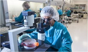
In recent times, PDX models or patient-derived xenograft models are getting heavily utilized in head and neck oncology research, specifically for studies on radiation response and organoids.
What is a Patient-Derived Xenograft?
Patient-derived xenografts (PDX) are xenograft models that have been derived from patient tumor tissues but never adapted to grow like cell line-derived xenografts. Patient-derived xenografts are beneficial than conventional cell line xenografts as they retain and maintain the genetic features of the native tumor besides mimicking the heterogeneity of the tumor microenvironment. PDX has many applications in drug efficacy tests, preclinical and clinical studies, and oncogenic biomarker discovery.
PDX Models in Head and Neck Cancer Research
Patient-derived xenografts have been developed from a wide range of head and neck tumor types like HPV-positive head and neck squamous cell carcinoma, HPV-negative head and neck squamous cell carcinoma, rare histology-based tumors as adenoid cystic carcinoma, and midline NUT carcinoma. The lab developed patient-derived xenograft can also be used for predicting patient response to the targeted drug agents, allowing clinically relevant studies.
The benefit of using PDX models for radiation response cancer studies is the use of human tumors rather than animal tumor models as there are significant differences between human and animal tumor cells in their fundamental regulatory response to radiation. The levels of DNA damage response proteins also differ between species and in human tumors, checkpoint activation is more efficient. Therefore, using human tumors is the more preferred method for pre-clinical studies and patient-derived xenografts have paved the path for it with much efficacy. These xenograft models also provide a fantastic native setting for conducting chemotherapy studies as they mimic body environment and physiology.
Another key application of these xenografts is the availability of organoid model regeneration in head and neck cancer research. Tumor organoids are directly derived from the patient tissue or from patient-derived xenograft models. The major limitations of using tissues of patients are ease of access, strict ethical regulations, and non-renewable tissue source for repeating research assays. Tumor organoid generation bypasses these limitations as these models provide expanded availability of cancer cells and an enhanced translation from in vitro organoids to in vivo xenografts. The tumor organoids which are derived from head and neck cancer patients mirror molecular, morphological, and genetic characteristics of the native tumor.
Limitations of PDX Models in Head and Neck Cancer Studies
The major limitations of the patient-derived xenograft model can be briefly put as:
- As these xenograft models are usually used with immune-compromised mice, it doesn’t provide a platform for testing the immune-oncology molecules and targets. These limitations can be overcome by using patient-derived xenografts with humanized mouse models.
- The xenografts are often established from the tissues which have been surgically removed while operating on a tumor and that brings a limitation of being unaware of how the surgically removed tissue will function similar to the patient body.
- Multiple passages have the possibility to cause potential biological changes and stromal loss in patient-derived xenografts but this can be overcome by using lower passage numbers and banking the models under optimized conditions.
- Specific limitations for head and neck cancers include engraftment rate based on HPV-negative / HPV-positive tumors as negative ones engraft slightly better than positive tumors.
To achieve high-end advancements in the field of head and neck cancer research, patient-derived xenograft models have proven to be clinically relevant preclinical models that pose several advantages over conventional in vivo models as they also pave the path towards regenerating 3D tumor organoids for higher research efficacy.
