According to the World Health Organization (WHO), cardiovascular diseases (CVDs) refer to a group of diseases that target the heart and blood vessels such as coronary heart disease and rheumatic heart disease. These diseases rank number one in deaths across the world- around 17.9 million lives each year! The WHO also lists that out of 5 deaths due to CVDs, 4 are accounted for by heart attacks and strokes with 1/3rd of these deaths in people below 70 years of age. The Centers for Disease Prevention and Control lists that one person in the U.S. loses their life due to cardiovascular disease every 37 seconds!
These statistics over the years have led to lots of research to understand the functioning and disease pathways of cardiomyocytes. This can also allow the designing of therapies to address CVDs. Primary cultures of cardiomyocytes have been obtained from mouse, rat, cat, rabbit and humans for short spans of time.
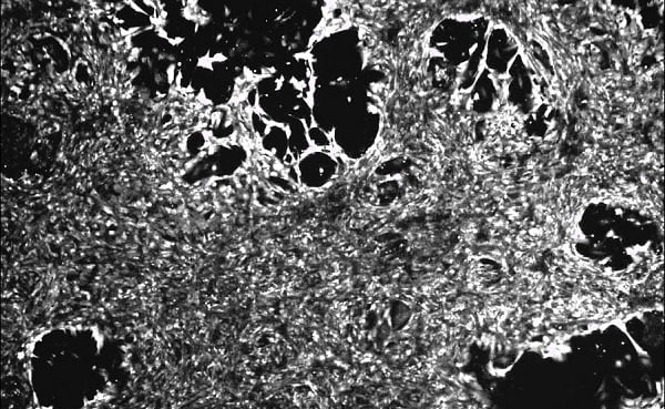
Primary cultures of cardiomyocytes have been reported to last for 2-3 weeks. The number of cells isolated from postnatal hearts is few as these cells do not usually proliferate in culture. The use of neonatal cardiomyocytes faces the challenge of loss of their contractile nature in few weeks of culturing. This is because the heart contains non-contractile cells such as endothelial cells, fibroblasts, and smooth muscle cells in addition to the contractile cardiomyocytes. Among these cells, the growth of fibroblasts is rapid can beat the growth of cardiomyocytes and even impede the contractions of the cells or cause dedifferentiation. This requires that primary cardiomyocyte cultures from heart samples are free from other cells.
Several approaches have been attempted in order to enrich cardiomyocytes in culture. One approach is the use of a pre-plating step. This is because non- cardiomyocyte cells can attach more quickly to culture plates than cardiomyocytes. Another system is the use of chemicals that target proliferation in the culture media such as cytosine-β-d-arabinofuranoside and 5-bromodeoxyuridine. These molecules are however cytotoxic and are potential mutagens. There is also the use of Percoll gradients that separate the non- cardiomyocytes of lower density from cardiomyocyte. Yet, this system too is limited to completely do away with non-cardiomyocytes especially, fibroblasts!
An article published by Phong and a team in the American Journal of Physiology-Cell Physiology discussed the primary culture of cardiomyocytes used tetramethylrhodamine methyl ester perchlorate (TMRM): a fluorescent stain that labels the mitochondria to enrich the cardiomyocytes. The cells that took up the stain at high amounts were cultured to establish viable neonatal rat cardiomyocyte. The cells could maintain spontaneous contractility and display responses to chronotropic agents (showing the expression of functional adrenergic and muscarinic receptors) and calcium cycling.
Such primary cultures of cardiomyocyte can allow for studying the process of development of these cells. They can also allow for the assessment of the toxicity of potential compounds used for treating CVDs. Such an assessment can be carried out at genomic and electrophysiological levels. They also offer exciting avenues to look at cell-based therapies of cardiomyocytes with stem cells!
References:
https://www.who.int/health-topics/cardiovascular-diseases/#tab=tab_1
https://www.cdc.gov/heartdisease/facts.htm
Phong D. Nguyen, Sarah T. Hsiao, PriyadharshiniSivakumaran, Shiang Y. Lim, and Rodney J. Dilley. Enrichment of neonatal rat cardiomyocytes in primary culture facilitates long-term maintenance of contractility in vitro. American Journal of Physiology-Cell Physiology 2012 303:12, C1220-C1228.
