
Hematopoietic stem cells (HSCs) are popular for their self-renewal potency. They are multipotent in nature, that is, they can transform into few lineages. They compose the blood and immune cell lineages. Their transplantation has numerous clinical applications in malignant and non-malignant disorders. As a standard line of treatment, HSC transplant has been a well-studied procedure. The applications of these cells have prompted deep research into the signaling mechanisms of their stem cell potency and modifications in their treatment procedure to ensure safe and effective therapy.
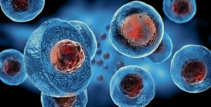
Human Bone Marrow Derived Hematopoietic Stem Cells
Hematopoietic stem cell was the first identified stem cell with self-renewal and differentiation potential. Their niche in the bone marrow comprises stromal and vascular components. They differentiate into common lymphoid and common myeloid progenitors (Fig 1). Common myeloid progenitors form megakaryocytes, erythrocytes, granulocytes, and macrophages. Common lymphoid progenitors form lymphocytes and natural killer cells. HSCs can be short-term or long-term depending on their bone marrow reconstitution ability. Long-term stem cells can replenish a lethally irradiated bone marrow, whereas short-term stem cells have limited self-renewal capacity.

Research has shown functional decline of HSCs with age. Their self-renewal capacity in older individuals is less than that of younger individuals. Research has proposed telomere shortening with each consecutive cell cycle and accumulation of DNA damage with every cell division as one of the possible reasons. But a limited number of cell divisions in HSCs have casted doubts on this theory. Additionally, scientists have noticed an asymmetric distribution of proteins associated with aging HSCs and caused by increased activity of CDC42. Inhibition of CDC42 resulted in symmetric protein distribution and improved HSC functioning. Another theory states that mutations accumulate in mitochondria with age and a dysfunctional mitochondria produces reactive oxygen species, which could cause the reduced functional capacity. Moreover, HSCs also demonstrate loss or alteration of epigenetic markers during cell division. The daughter cells with modified epigenetic markers proliferate by clonal hematopoiesis, affecting the self-renewal potential.
Isolation of Hematopoietic Stem Cells
HSC Extraction from Bone Marrow: Bone marrow is predominantly present in the long bones. The extraction procedure of HSCs from bone marrow involves inserting a needle into the iliac crest or the hip bone and aspirating the bone marrow fluid. Another technique to extract HSCs is to mobilize them from bone marrow into peripheral blood by administration of granulocyte-colony stimulating factor (GCSF) . A separator machine isolates the mononuclear cells and HSCs from the peripheral blood. This technique is termed as apheresis and is painless as compared to the former technique.
HSC Isolation: HSCs are isolated based on their markers. CD34 is the key identification marker for human HSCs. Other markers are CD90+ c-kit-/low Lin– CD38– CD45RA–. The isolation process requires treatment of cell suspension with antibodies specific to these markers and separation by magnetic-activated cell sorting (MACS) or fluorescence-activated cell sorting (FACS), depending on the label on the antibody.
Cell Sorting: MACS is employed for the isolation process for antibodies labeled with iron oxide beads. The application of a magnetic field on the cell suspension retains antibody bound HSCs while eluting the rest of the cells, providing a pure population of HSCs.
In case the antibody is labeled with fluorescent dyes like FITC or PE, then cells are separated by FACS . The cell suspension flows through the instrument where hydrodynamic focusing ensures the extrusion of one cell from the nozzle. The laser excitation captured the fluorescence signal of each cell. Based on the signal, an electric ring adds a specific charge on the cell. Afterwards, an electrostatic system deflects the cell droplets into different containers based on their charge. This sorting process is especially beneficial for the separation of more than one population.
Staining: Staining with Hoechst 33342 and rhodamine 123 dyes can also separate HSCs from other cells. These dyes dimly stain the metabolically inactive cells. But HSCs also show dim staining with these dyes due to their efflux by HSC membrane transporters.
Functional Characterization of Hematopoietic Stem Cells
During the isolation process, HSCs are characterized based on their surface markers. But the following assays evaluate their self-renewal and differentiation capacity.
Colony Forming Unit (CFU) Assay: The assay assesses the differentiation potential of HSCs. The assay entails mixing the isolated HSCs in a semisolid media- methylcellulose and pouring it evenly into a 35 mm dish alongside another 35 mm dish containing sterile water without the lid for maintaining humidity. Place both the dishes in a 100 mm dish in an incubator and count the colonies after 2 weeks under a microscope. CFU-E, i.e., colony-forming unit of erythroblasts, shows a red or pinkish color and forms one or two clusters comprising 8-200 erythroblasts. CFU-G is the colony-forming unit containing a homogenous population of granulocytes, CFU-GM is the colony-forming unit consisting of both granulocytes and macrophages, whereas CFU-GEMM is the colony-forming unit comprising granulocytes, erythrocytes, macrophages, and megakaryocytes.
Long-Term Culture Initiating (LTC-IC) Assay: This assay measures whether isolated HSCs can initiate and sustain renewal and differentiation potential for a long time period. The assay follows coculture of HSCs over a feeder layer of fibroblast cells or immortalized stromal cell lines and performing colony-forming unit assay on the non-adherent cells after 5 weeks. After 5 weeks, non-adherent cells are collected and colony forming unit assay is repeated on them.
Cobblestone Area-Forming Cell (CAFC) Assay: This assay is a variation of LTC-IC assay. It also involves co-culture of HSCs on a stromal layer. Within a week, dark cobblestone-shaped regions appear beneath the stromal layer, indicative of cell proliferation
Applications of Human Bone Marrow Derived HSCs
HSCs transplant is also termed as bone marrow transplant. It requires replacing diseased HSCs with healthy HSCs. Since the 1950s, HSC transplantation has been optimized with respect to the HSC source of, their extraction process, dosage, and additional medications required during the transplant.
Genetic Disorders: HSCs form the blood cell lineages. Any genetic aberrations in these cells result in defective terminal lineages, causing severe disorders such as severe combined immunodeficiency disease (SCID), sickle cell anemia, thalassemia, etc. The life expectancy significantly declines in these disorders and mandates the transplant of healthy HSCs to provide a healthy population of blood cell lineages.
Hematological Malignancies: Several hematological malignancies, such as multiple myeloma, Non-Hodgkin’s lymphoma, and acute myelogenous leukemia, arise from genetically mutated precursors. Chemotherapy and radiation eliminate the malignant cells, but a person still requires healthy cells to maintain the normal functions. Transplantation of healthy HSCs after chemotherapy restores the healthy cell population.
Radiation Poisoning: HSC transplant was the first line of treatment to rescue the radiation-exposed victims of war. Lethal radiation severely diminishes the stem cell population, leaving a limited number of differentiated cells. HSC transplant can reconstitute the bone marrow population of healthy stem cells.
Product-Related Queries, Or Partnership Inquiries
Conclusion
HSC transplantation is a widely used procedure in clinical settings. It is the end-stage treatment option for several disorders. Scientists are still conducting research on HSCs regarding their signaling pathways, potency, and improved treatment outcomes. Kosheeka provides human bone marrow-derived CD34+ HSCs. A team of trained professionals rigorously evaluate the cells for their functional potential, viability, and quality. We also offer supporting media for the culture of HSCs.
FAQs
What are the different assays for evaluating the regeneration potential of HSCs?
Colony forming unit (CFU) assay, LTC-IC assay and CAFC assay are used to assess the proliferation and differentiation potential of HSCs.
What are the effects of aging on HSCs?
Aging has been seen to reduce the self-renewal potential of HSCs. Several causative factors have been outlined for age-induced effects on HSCs but the mechanism is poorly understood.
What is the difference between long-term and short-term HSCs?
Long-term HSCs have higher self-renewal capacity as compared to short-term HSCs, and they could reconstitute the irradiated bone marrow.
What does the CFU assay measure?
CFU assay analyzes the differentiation potential of HSCs in different lineages.
