

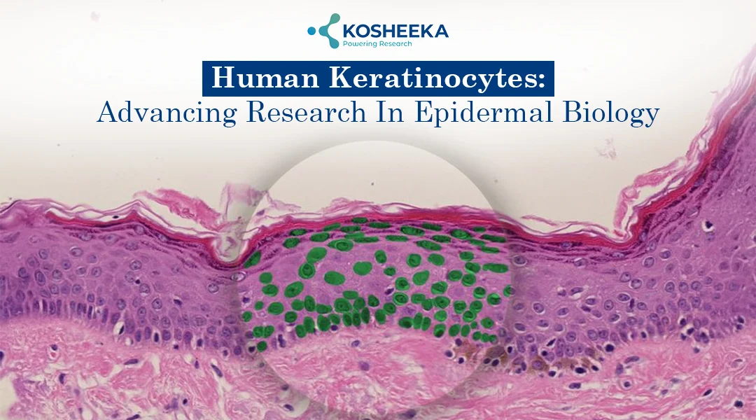
Research with human skin in the laboratory involves the use of human keratinocytes. Skin is made up of different cell types that work together. The detailed understanding involves researchers’ efforts to grow cells under controlled laboratory conditions. Keratinocytes are a natural starting point; they make up most of the epidermis and are directly involved in maintaining its structure and function.
Human keratinocytes are widely investigated in skin research, including cell growth, differentiation, understanding barrier function, and response to the external environment. Detailed understanding of these cells’ behaviour in culture reveals useful information related to normal epidermal biology and skin disorders model. Keratinocyte stem cells are of particular interest because they help maintain the epidermis by producing new cells throughout life.
It is crucial to maintain adequate keratinocyte culture conditions. This involves considering cell origin, culture medium, macro and micronutrient availability, passage number, differentiation, etc. The current article focuses on unveiling key features of Human Keratinocyte Culture and their role in various biomedical research.
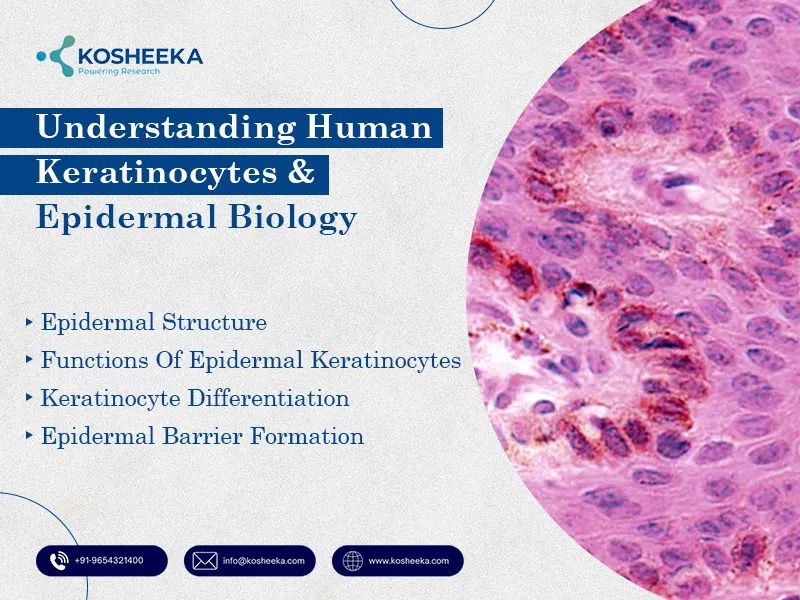
Understanding Human Keratinocytes and Epidermal Biology
Human keratinocytes are predominant epidermal cells that make up the skin’s outermost layer. The role of epidermal keratinocytes extends beyond the development of structural support, maintenance of the skin barrier, combatting environmental stress, and regulation of epidermal tissue. Understanding the key biology is crucial. This involves:
Epidermal Structure
- Basal Layer (Stratum Basale): Deepest epidermal layer; keratinocytes proliferate. Consists of epidermal keratinocyte stem cells and progenitor cells. Aid in skin renewal
- Spiny Layer (Stratum Spinosum): Connects strong intercellular junctions known as desmosomes. The connection enables epidermal keratinocytes to withstand mechanical stress
- Granular Layer (Stratum Granulosum): Epidermal keratinocytes undergo differentiation. It accumulates various bioactive molecules like proteins and lipids involved in barrier development. Lamellar bodies are involved in releasing lipids
- Cornified Layer (Stratum Corneum): Outermost skin layer; this involves flattened, terminally differentiated keratinocytes (corneocytes). The cells are embedded with a lipid-rich matrix
Functions of Epidermal Keratinocytes
- Maintain Skin Integrity: Epidermal Keratinocytes produce various structural proteins (keratins). This supports epidermal strength. It enables skin to withstand external stress, everyday friction and mechanical stress
- Support Epidermal Renewal: Basal keratinocytes replace the lost cells via normal shedding. This feature supports cellular turnover
- Immune Response: Keratinocytes recognise environmental foreign particles or microbial signals. The cells release various cytokines and chemokines that invoke the immune response and recruit local inflammatory molecules
- Injury Response: On skin damage, human keratinocytes migrate and proliferate to the injury site. They enable epidermal surface renewal and wound repair
Keratinocyte Differentiation
- Proliferation to Specialisation: Basal keratinocytes proliferate and move upward to the epidermal layer. They acquire specialized structural and functional properties
- Protein Expression: Distinct keratinocytes release distinct proteins. Basal keratinocytes (keratins K5 and K14) and suprabasal keratinocytes (K1 and K10). At the maturity stage, involucrin, filaggrin, loricrin, etc., are released
- Differentiation: Keratinocyte differentiation is influenced by factors including extracellular calcium concentration. They are major contributors supporting proliferation and differentiation. The differentiation markers and cellular morphology reflect cell health
Epidermal Barrier Formation
- Cornified Envelope Formation: Develops into a specialized protein-rich envelope; it strengthens the outer epidermal layer
- Barrier Lipids: Ceramides, cholesterol, fatty acids, etc. form the ECM matrix
- Tight Junction Formation: Granular layer helps with substance movement through the epidermal layer
Keratinocyte Stem Cells: How is Epidermal Renewal Maintained?
All forms of keratinocytes have distinct functionality. Their roles differ depending on their stage of development and ability to produce new cells.
Key Function
- Epidermal Keratinocyte Stem Cells: These long-lived cells can renew themselves and generate cells that contribute to epidermal maintenance. They are found in the basal epidermis and specialised regions of hair follicles.
- Progenitor Cells: These cells have a more limited capacity for self-renewal and produce keratinocytes that support routine tissue maintenance.
- Transit-Amplifying Keratinocytes: These cells divide several times before their descendants progress towards differentiation, helping increase the number of cells available for epidermal renewal.
Self-Renewal and Tissue Homeostasis
- Cell Supply Maintenance: Stem cells efficiently preserve cell population have renewal capacity
- Epidermis Balance: Cell production maintains coordination with the loss of cells from the skin surface to maintain normal epidermal structure.
- Responding to Injury: When the skin is damaged, keratinocyte populations can adjust their activity to support tissue repair.
How Keratinocytes are Cultured: Methods and Key Considerations
Growing research interest involves the use of human keratinocytes. This involves consideration of cell source, culture conditions, and handling practices. All these factors influence the way the cells grow and behave. The key keratinocyte culture conditions include:
Primary Human Keratinocytes vs. Immortalised Cell Lines
- Primary Human Keratinocytes: Isolated directly from human tissue. These cells closely represent epidermal physiology. The growth capacity of the cells is limited. However, variation in donors can influence the final research outcome.
- Immortalised Cell Lines: Keratinocyte secondary cell culture involves modified cells. Cell lines (HaCaT) are widely used for large-scale research. The cell lines are vital in developing preliminary studies, obtaining experimental replicates, high-throughput techniques, etc. These cells have a longer lifespan and can have multiple passages
- Adequate Cell Model Selection: Choosing an adequate keratinocyte culture depends on the research question or goals
Culture Medium
- Culture Media and Supplements: Keratinocytes require an appropriate nutrient-rich medium, often supplemented with growth factors such as EGF. Calcium concentration also matters, as it can influence cell adhesion and differentiation.
- Culture Substrates: Standard culture surfaces work for many studies, while extracellular matrix coatings may be useful when specific cell–matrix interactions are being investigated.
- Matching Conditions to the Study: Conditions that support cell proliferation may differ from those used to study differentiation or epidermal barrier formation.
Culture Condition
- Passage Number: Primary keratinocytes can change as they are repeatedly subcultured. Keeping track of passage history helps researchers identify potential sources of variation.
- Cell Confluence: How densely cells grow can affect their behaviour, so seeding density and subculture timing should remain consistent.
- Differentiation Control: Maintenance of culture conditions (availability of calcium levels and growth supplements) can influence how keratinocytes mature. Cell morphology and markers such as K14 and K10 can help researchers assess their differentiation status.
*NOTE: Researchers must use consistent culture protocols, document changes in cell behaviour, and select favourable culture conditions. Kosheeka, India, is the leading manufacturer and supplier of various animal cell culture cells, including human keratinocytes. The product comes with a Certificate of Authentication (CoA)
What are the Key Research Applications?
- Understanding skin barrier biology, wound repair
- Dermatological disease modelling and inflammatory responses
- Screening of various dermatological molecules
- Toxicity assessment
- Development of 3D epidermal models or organoid culture
Conclusion
Human Keratinocyte Culture is an effective in vitro model for various skin and biomedical research. These are primary cells that form the majority of the epidermal skin layer. The key mechanisms involve maintenance of structure, the skin barrier, protein production, protection against foreign molecule exposure, etc. Researchers procuring human keratinocyte cells for research must ensure they meet superior standards.
FAQ’s
Q- What are human keratinocytes?
Human keratinocytes form the outermost epidermal skin layer. They are responsible for forming protective barriers. Keratinocytes are involved in skin renewal, tissue repair, and immune response. Human keratinocytes enable researchers to understand skin functions, what happens in dermatological conditions, and how topical ingredients affect skin cells under controlled laboratory conditions.
Q- How do keratinocyte stem cells differ from other epidermal keratinocytes?
Keratinocyte stem cells can self-renew and generate progenitor cells that contribute to epidermal maintenance and renewal. Other keratinocytes progressively differentiate to form the specialised layers of the epidermis. Understanding these differences helps researchers investigate tissue homeostasis, skin regeneration, and cellular differentiation.
Q- What factors are important for successful keratinocyte cell culture?
Keratinocyte cell culture requires the presence of controlled laboratory conditions. This involves the presence of suitable culture media, growth supplements, cell seeding density, etc. Researchers should also monitor cell morphology, viability, contamination, and differentiation status to maintain reliable and reproducible experimental results.
Q- What are the key research applications?
Human keratinocyte culture has a wide range of research applications. This includes skin barrier function and wound repair, dermatological disease modelling and topical ingredient evaluation. Keratinocytes are also used to reconstruct epidermal models and for tissue engineering research.

Author: Dr. Siuli Shaw
PhD in Biotechnology | Scientific Writer | Advancells Group
Translating cutting-edge science into impactful scientific communication.
Research Interests: Cancer Therapeutics | Nanotechnology | Stem Cell Research | Regenerative Medicine


